
Diastasis Rectus Abdominis: Assessment and Progressive Loading
Learn to assess diastasis rectus abdominis and use progressive abdominal loading to restore strength, function and exercise confidence.
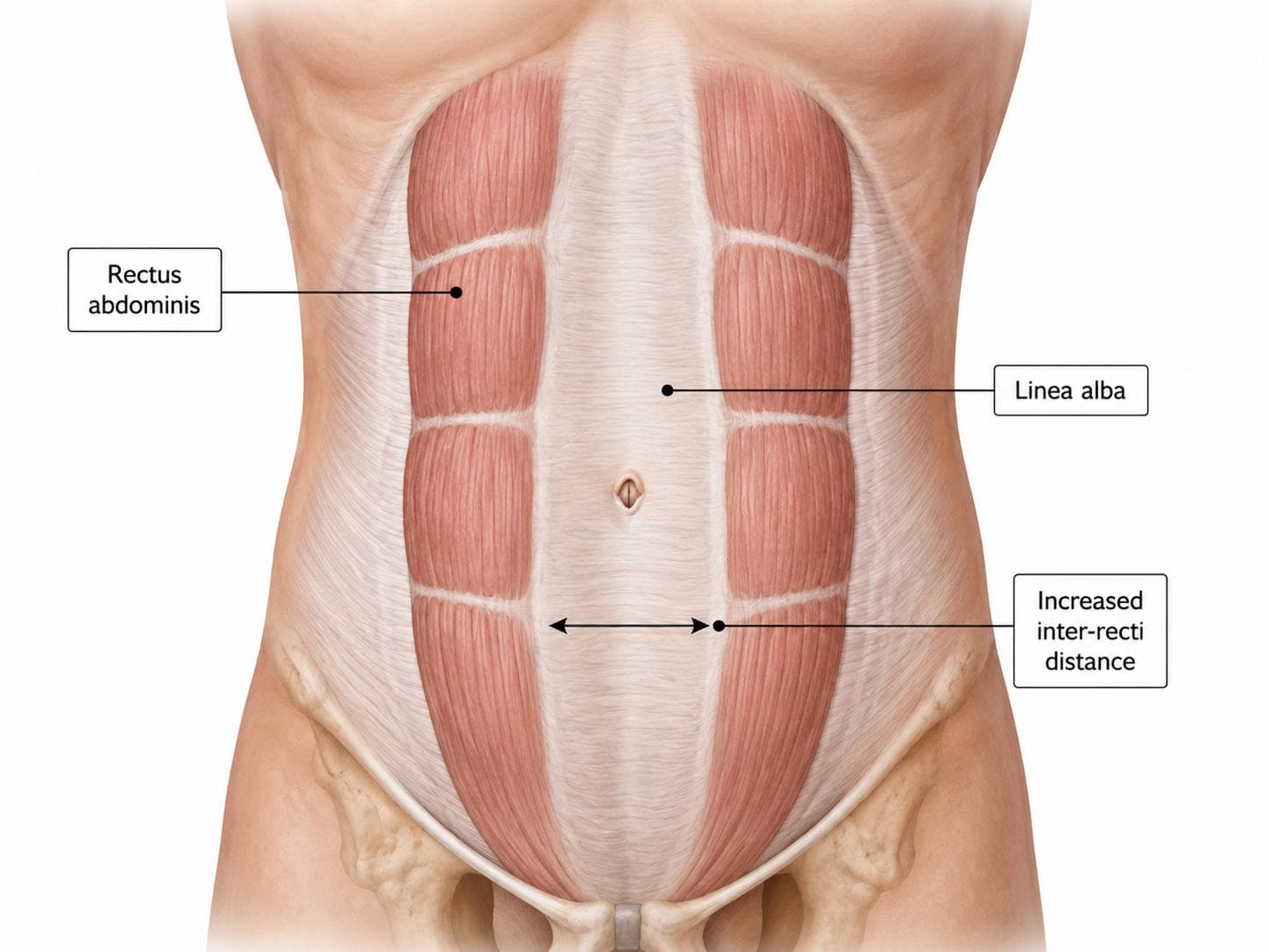
Diastasis rectus abdominis is frequently discussed as a “gap” between the abdominal muscles. Clinically, however, rehabilitation should not focus only on reducing this distance. The physiotherapist must also assess how the abdominal wall generates tension, manages load and supports functional activities such as rolling, lifting, exercising and caring for an infant.
Current evidence supports structured abdominal exercise, but no single exercise programme has been established as universally superior. Treatment should therefore follow a progressive, criteria-based approach rather than a fixed list of “safe” and “unsafe” exercises.
What Is Diastasis Rectus Abdominis?
Diastasis rectus abdominis, or DRA, refers to widening of the linea alba with increased separation between the medial borders of the two rectus abdominis muscles. The European Hernia Society defines rectus diastasis as a separation wider than 2 cm, although the measured distance must always be interpreted alongside symptoms and function (Hernández-Granados et al., 2021).
DRA commonly develops during pregnancy as the abdominal wall adapts to the enlarging uterus. It may persist after childbirth, but it can also occur in men and non-pregnant women in association with obesity, repeated increases in abdominal pressure, connective-tissue characteristics or previous abdominal surgery.
Unlike a ventral hernia, DRA does not involve a true defect or hole in the abdominal fascia. However, a patient may have both rectus diastasis and an umbilical or epigastric hernia, making differential screening essential.
The patient may report:
- A visible midline bulge during movement
- Reduced confidence in the abdominal wall
- Difficulty with lifting, transfers or exercise
- Abdominal weakness or fatigue
- Cosmetic or body-image concerns
- Associated low-back, pelvic-girdle or pelvic-floor symptoms
These symptoms are not determined by inter-recti distance alone. Some people with a wide separation function well, whereas others with a smaller distance experience marked weakness or difficulty controlling pressure.
Why Assessment Must Go Beyond Gap Width
Inter-recti distance, or IRD, describes the measurable distance between the two rectus abdominis muscles. It is useful for documenting abdominal-wall anatomy, but it does not provide a complete measure of rehabilitation success.
A 2026 meta-analysis of nine randomised trials involving 450 participants found that structured exercise produced a greater reduction in IRD than no intervention or standard care. However, this anatomical change did not produce a corresponding improvement in disability measured using the Oswestry Disability Index (Capoccia Giovannini et al., 2026).
This distinction is clinically important. The aim of physiotherapy is not simply to “close the gap". Rehabilitation should improve the following:
- Abdominal muscle strength and endurance
- Tension transfer through the linea alba
- Breathing and pressure-management strategies
- Lumbopelvic control
- Confidence during daily and recreational activities
- Capacity to lift, carry, run or return to sport
Research also challenges the traditional belief that all curl-ups or planks must be avoided. In a randomised trial, a 12-week programme containing head lifts and curl-ups improved rectus abdominis strength and thickness without worsening IRD, pain or pelvic-floor symptoms (Gluppe et al., 2023).
Exercises should therefore be selected according to the patient’s capacity and response, not their name alone.
Clinical Assessment of Diastasis Rectus Abdominis
Assessment begins with the patient’s history. Relevant information includes pregnancy and delivery history, postpartum stage, abdominal or pelvic surgery, exercise background, pain, urinary or faecal symptoms, vaginal heaviness, constipation, lifting demands and the activities the patient wants to resume.
The physiotherapist should ask what concerns the patient most. For one person, the primary problem may be difficulty lifting a child. For another, it may be abdominal bulging during exercise or reduced confidence returning to the gym.

Observation and palpation
The abdominal wall may be observed at rest and during tasks such as the following:
- Head lift or modified curl-up
- Rolling and rising from bed
- Sit-to-stand
- Coughing
- Squatting or lifting
- Quadruped or plank positions
The examiner notes the location and extent of separation, visible doming, breath-holding, rib flare and the patient’s ability to maintain abdominal-wall tension.
Finger-width palpation is accessible and useful for initial screening, but finger dimensions vary between examiners. Digital callipers and diastometers provide more numerical information, while ultrasound imaging offers the most objective assessment of IRD when accurate measurement or monitoring is required. Whichever method is used, the position, measurement site and activity should be standardised at reassessment (Petronilla et al., 2023).
Assessing tension and function
The examiner should evaluate more than width. The linea alba may feel firm and tensioned during contraction, or soft and poorly tensioned despite a relatively small IRD. Depth of the separation, tissue behaviour and abdominal-wall control should therefore be documented.
Functional assessment may include:
- Pain-free rolling and transfers
- Repeated sit-to-stand
- Squat and hip-hinge control
- Lifting an object from floor to waist
- Carrying tasks
- Trunk endurance
- Single-leg loading
- Impact testing when return to running is a goal
Pelvic-floor symptoms, breathing mechanics, hip strength and general physical conditioning must also be assessed because DRA rarely exists as an isolated “rectus muscle” problem.
Principles of Progressive Abdominal Loading
Progressive loading exposes the abdominal wall to gradually increasing demands so that it can adapt. The programme should begin at a level the patient can control and progress towards the forces required in real life.
Early rehabilitation may emphasise breathing, coordination and low-load movement. These exercises are useful starting points, but they should not become permanent restrictions. The eventual goal is to restore the patient’s ability to tolerate resistance, speed, impact and functional lifting where appropriate.
Important principles include:
- Coordinate breathing with effort. Exhaling during the difficult phase can help a patient who initially holds their breath or generates excessive pressure. As capacity improves, normal breathing should be maintained during sustained and heavier tasks.
- Train the entire trunk. Rehabilitation should include the rectus abdominis, obliques, transversus abdominis, diaphragm, spinal muscles, pelvic floor and hip musculature rather than attempting to isolate one “deep core” muscle.
- Use symptoms to guide dosage. A temporary change in abdominal shape is not automatically harmful. However, marked uncontrolled bulging accompanied by pain, pelvic heaviness, leakage or loss of technique indicates that the current task may exceed the patient’s capacity.
- Progress load, not only exercise complexity. An exercise can be advanced through additional repetitions, resistance, lever length, range, speed, reduced support or greater functional relevance.

General therapeutic-exercise principles suggest beginning with a manageable volume and increasing demand as movement quality, recovery and tolerance improve (Kisner et al., 2023).
Four-Stage Progressive Loading Framework
The stages below are not fixed postpartum timelines. Entry and progression depend on healing, symptoms, baseline fitness and functional goals.

Motor-control exercises may initially be performed for one or two sets of six to eight slow repetitions. Strength exercises can later progress towards two or three sets of approximately six to twelve repetitions, two or three times per week. The exact dosage should reflect the patient’s recovery, sleep, activity level and training history.
The evidence does not identify one superior exercise category. Recent reviews suggest that several structured approaches—including conventional strengthening, deep-core programmes and hypopressive exercise—may reduce IRD, but study quality and protocols remain inconsistent (Bigdeli et al., 2025; Skoura et al., 2024; Soto-González et al., 2024).
Monitoring, Modification and Referral
The response during exercise and over the following 24 hours should guide progression.
Continue or progress when the patient demonstrates controlled breathing, acceptable abdominal-wall behaviour and no increase in pain or pelvic-floor symptoms. Maintain or slightly reduce the dose when technique deteriorates near the end of a set but symptoms settle quickly. Regress the range, resistance or lever length when the patient develops marked doming, breath-holding, pain, leakage, heaviness or next-day aggravation.

Temporary binders or taping may provide comfort and confidence for selected patients, but they should not replace progressive muscle loading.
Medical or surgical assessment is indicated when there is the following:
- A focal, painful or non-reducible abdominal bulge
- Suspected umbilical, epigastric or incisional hernia
- Severe or unexplained abdominal pain
- Nausea or vomiting associated with a bulge
- Postoperative wound complications
- Progressive neurological or systemic symptoms
- Persistent functional limitation despite appropriate rehabilitation
Surgical consultation may also be considered for significant ongoing symptoms, a concomitant hernia or major patient-reported functional and body-image concerns after conservative management. The European Hernia Society recommends that physiotherapy may be considered before surgical treatment (Hernández-Granados et al., 2021).
Viva-Ready Summary
Diastasis rectus abdominis is widening and thinning of the linea alba with increased separation between the rectus abdominis muscles. A separation wider than 2 cm is commonly used as a clinical definition, but gap width should not be assessed in isolation.
Assessment includes history, abdominal-wall observation, palpation or objective IRD measurement, linea alba tension, breathing, pelvic-floor symptoms and functional loading. Ultrasound is useful when precise or repeatable measurement is required.
Management begins with breathing and low-load trunk control before progressing to limb loading, multidirectional strengthening, functional resistance and higher-level activity. Curl-ups, planks and loaded exercises are not automatically contraindicated; they may be introduced when the patient can tolerate them without significant symptoms or loss of control.
One-line recall: Treat the function and load capacity of the abdominal wall—not merely the measured gap.
References
Bigdeli, N., Yalfani, A., Doosti-Irani, A., & Qodrati, A. (2025). An evidence-based comparison of rehabilitation strategies for diastasis recti abdominis in postpartum women: A systematic review and network meta-analysis. Scientific Reports, 15, 39591. https://doi.org/10.1038/s41598-025-22574-2
Capoccia Giovannini, S., Hoffmann, H., Bracale, U., et al. (2026). Non-operative management of postpartum diastasis recti: A systematic review and meta-analysis of randomised controlled trials. Hernia, 30, Article 164. https://doi.org/10.1007/s10029-026-03671-1
Gluppe, S. B., Ellström Engh, M., & Bø, K. (2023). Curl-up exercises improve abdominal muscle strength without worsening inter-recti distance in women with diastasis recti abdominis postpartum: A randomised controlled trial. Journal of Physiotherapy, 69(3), 160–167. https://doi.org/10.1016/j.jphys.2023.05.017
Hernández-Granados, P., Henriksen, N. A., Berrevoet, F., Cuccurullo, D., López-Cano, M., Nienhuijs, S., Ross, D., & Montgomery, A. (2021). European Hernia Society guidelines on management of rectus diastasis. British Journal of Surgery, 108(10), 1189–1191. https://doi.org/10.1093/bjs/znab128
Kisner, C., Borstad, J., & Colby, L. A. (2023). Therapeutic exercise: Foundations and techniques (8th ed.). F. A. Davis.
Petronilla, O. C., Uruchi, E. A., Francis, E. C., & Linda, U. C. (2023). Assessment of diastasis recti abdominis: Clinimetric properties of four measuring instruments. International Journal of Health Sciences, 17(6), 6–14.
Skoura, A., Billis, E., Papanikolaou, D., Xergia, S., Tsarbou, C., Tsekoura, M., Kortianou, E., & Maroulis, I. (2024). Diastasis recti abdominis rehabilitation in the postpartum period: A scoping review of current clinical practice. International Urogynecology Journal, 35(2), 491–520. https://doi.org/10.1007/s00192-024-05727-1
Soto-González, M., Da Cuña-Carrera, I., Lantarón-Caeiro, E., & Pascoal, A. (2024). Effect of hypopressive and conventional abdominal exercises on postpartum diastasis recti: A randomised controlled trial. PLOS ONE, 19(12), e0314274. https://doi.org/10.1371/journal.pone.0314274


