
Introduction to Musculoskeletal Assessment: A Structured Approach
Learn a universal musculoskeletal assessment sequence using subjective examination, observation, AROM, PROM, resisted tests, special tests, palpation and neurovascular screening.
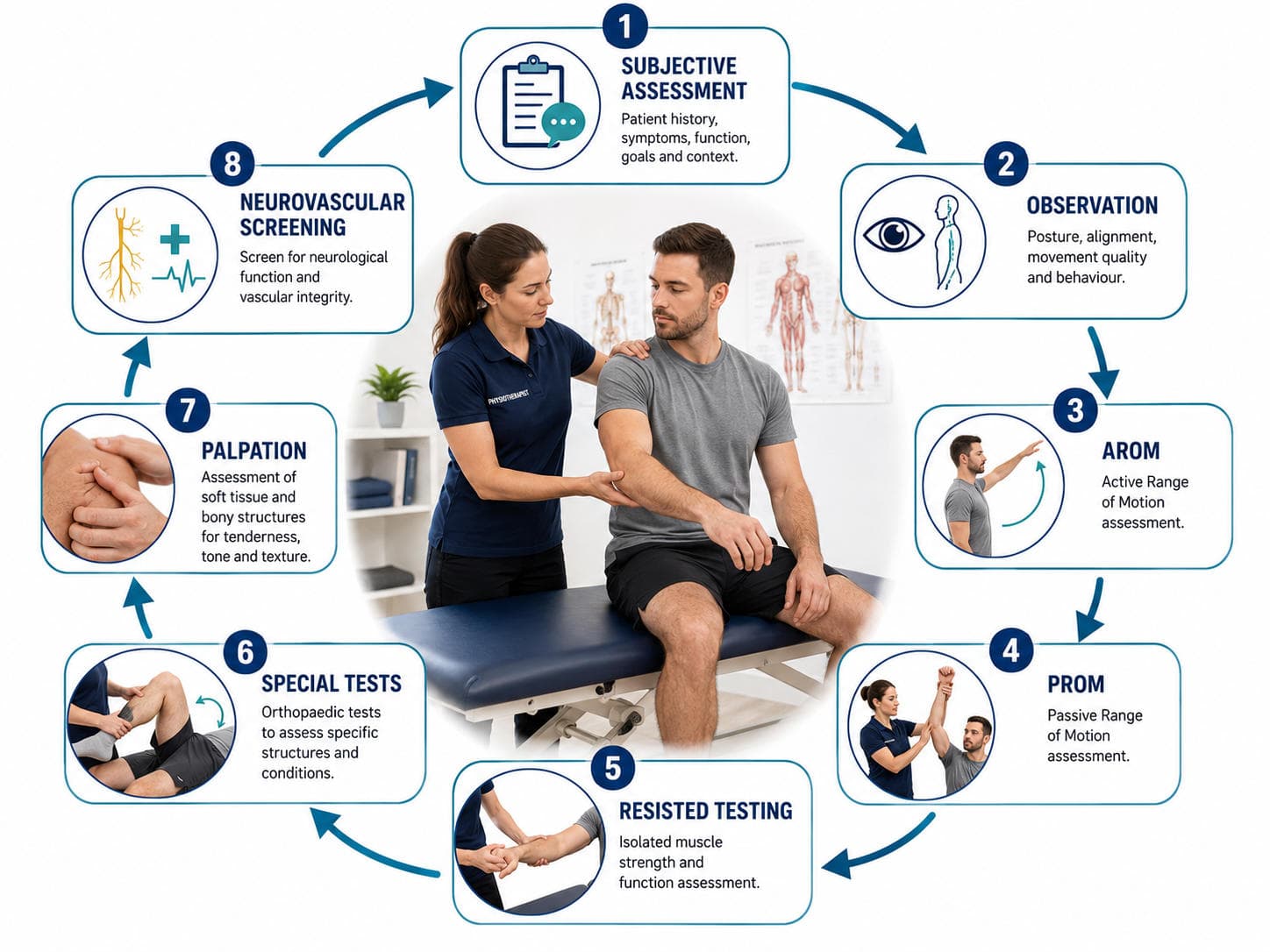
Musculoskeletal assessment is not a collection of unrelated special tests. It is a progressive clinical reasoning process in which each stage confirms, modifies or rejects the hypotheses developed during the previous stage. A reliable sequence for most peripheral joints is the following:
Subjective assessment → observation → active range of motion → passive range of motion → resisted testing → special tests → palpation → neurovascular screening
This framework can be adapted to the shoulder, elbow, wrist, hip, knee, ankle or another musculoskeletal region. It also gives students a predictable structure during practical examinations while ensuring that safety, function and differential diagnosis are not overlooked (Magee & Manske, 2021; Ryder & Barnard, 2023).
Why a Structured Assessment Sequence Works
The purpose of assessment is not merely to name an injured structure. It is to determine:
- whether the presentation is suitable for physiotherapy;
- which activities and movements reproduce the problem;
- whether the dominant impairment is related to mobility, contractile tissue, neurological function or another system;
- how the condition affects function and participation; and
- which findings can be reassessed after treatment.
The sequence should begin broadly and become progressively more specific. The subjective assessment establishes possible diagnoses and safety concerns. Observation and movement testing then identify the patient’s main functional and physical impairments. Resisted tests, selected special tests and palpation provide additional evidence, while neurovascular screening helps exclude nerve or vascular involvement.
This reflects contemporary best-practice musculoskeletal care, which emphasises patient-centred assessment, red-flag screening, psychosocial factors, physical examination and selective rather than routine use of imaging (Lin et al., 2020).
Subjective Assessment: Build the Initial Hypothesis
The subjective examination directs everything that follows. Begin by recording the patient’s presenting complaint in their own words and identifying what they want to regain—for example, overhead reaching, walking, stair climbing, work, sleep or sport.
Clarify the site, nature, intensity and behaviour of symptoms. Ask about onset, mechanism of injury, duration, progression, aggravating factors, easing factors, 24-hour behaviour and previous episodes. A traumatic twisting injury creates different hypotheses from gradual pain associated with repetitive loading.
Functional questions are especially important. Ask what the patient cannot do, what they can still do and whether performance is limited by pain, weakness, stiffness, instability, fear or fatigue. Medical history, medication, imaging, previous treatment, occupation, sport and dominant side may change both the diagnosis and management plan.
Screen for neurological symptoms such as numbness, paraesthesia, radiating pain, weakness, altered coordination or changes in bladder and bowel function where relevant. Also consider psychosocial or “yellow-flag” factors such as fear of movement, catastrophising, low recovery expectations, sleep disturbance and work-related concerns.
Before proceeding, identify features suggesting fracture, infection, tumour, inflammatory disease, visceral referral, progressive neurological compromise or vascular pathology. Red flags should be interpreted collectively within the patient’s clinical context rather than as isolated yes-or-no items (Storari et al., 2025).
Clinically accurate answer:
“From the subjective assessment, my initial hypotheses are ____. I will first observe the patient and then use movement testing to determine whether the dominant problem is contractile, articular, neurological or referred.”
3. Objective Assessment: Observation, AROM, PROM and Resisted Tests
Before touching the patient, explain the procedure, obtain consent, expose the region appropriately and compare with the unaffected side where relevant.

Observation
Observe the patient globally before focusing on the symptomatic region. Note their gait, transfers, resting posture and willingness to use the affected limb. Local inspection may reveal swelling, redness, deformity, muscle atrophy, bruising or surgical scars.
Observation should generate questions rather than conclusions. For example, scapular asymmetry does not independently diagnose shoulder pathology, but it may prompt closer assessment of movement control and muscle performance.
Active range of motion
Ask the patient to perform the movement independently. Record:
- available range;
- location and timing of pain;
- movement quality;
- compensatory movements;
- apprehension or instability; and
- whether the patient’s familiar symptoms are reproduced.
Begin with the least painful movement and examine the most provocative movement later. Functional movements such as a squat, step-down, hand-behind-back movement or overhead reach may be included before isolated joint testing.
Passive range of motion
The examiner then moves the relaxed limb through the available range. Compare active and passive findings.
A large difference between limited AROM and relatively preserved PROM may suggest pain inhibition, weakness, tendon dysfunction, neurological involvement or impaired motor control. A similar limitation of AROM and PROM may suggest joint, capsular, degenerative or highly irritable pathology. These are reasoning patterns, not stand-alone diagnoses.
Assess the end feel only when safe. Avoid forceful overpressure following significant trauma, when fracture or dislocation is suspected, or when symptoms are highly irritable.
Resisted isometric testing
Position the joint so that the target tissue can be tested safely without excessive joint movement. Apply resistance gradually and compare pain and strength with the opposite side.
A painful but reasonably strong contraction may support a contractile-tissue hypothesis. Marked weakness may reflect pain inhibition, major tissue disruption, neurological impairment or poor effort. Therefore, resisted testing must be interpreted alongside the history, range-of-motion findings and functional performance rather than in isolation (Magee & Manske, 2021).

4. Special Tests, Palpation and the Neurovascular Screen
Special tests should be selected after the history and basic movement examination have narrowed the differential diagnosis. Performing every test remembered for a region wastes time, irritates symptoms and produces findings that are difficult to interpret.
Use tests that answer a specific question:
“I suspect ____. I will perform ____ because it may increase or decrease the probability of that hypothesis.”
A positive test should reproduce the relevant symptom or demonstrate the intended finding—not simply cause nonspecific discomfort. Evidence indicates that many individual orthopaedic tests have limited diagnostic performance; they are most useful when interpreted with the history, movement pattern and other examination findings (Gismervik et al., 2017).
Palpation follows targeted testing. Assess temperature, swelling, tenderness, tissue continuity, bony landmarks and local muscle tone. Local tenderness can support a hypothesis but rarely identifies the diagnosis independently.
Complete a region-appropriate neurovascular screen. This may include:
- dermatomes, myotomes and reflexes;
- peripheral nerve sensation and motor testing;
- distal pulses and capillary refill;
- skin temperature and colour; and
- comparison between sides.
A detailed screen is particularly important when symptoms radiate, paraesthesia or weakness is reported, trauma has occurred, distal circulation appears altered or the symptoms cannot be explained by local tissue testing.
5. Worked Example: Suspected Rotator-Cuff-Related Shoulder Pain
A 42-year-old teacher reports gradual lateral shoulder pain for six weeks, aggravated by overhead writing, dressing and lying on the affected side. There was no trauma, systemic illness or paraesthesia.
Subjective hypothesis: Rotator-cuff-related shoulder pain is considered, while cervical referral, adhesive capsulitis, significant cuff tear and acromioclavicular pathology remain differential diagnoses.
Observation: There is no deformity, swelling or obvious muscle wasting. The patient uses mild scapular elevation when raising the arm.
AROM: Shoulder abduction reproduces familiar pain through the middle portion of elevation. Flexion is mildly painful, while cervical movements do not reproduce the shoulder symptoms.
PROM: Passive shoulder movement is almost full, with mild pain near end-range elevation. The relative preservation of PROM makes a marked capsular restriction less likely.
Resisted testing: Resisted abduction and external rotation reproduce pain, but strength is only mildly reduced. This supports a load-sensitive contractile-tissue presentation rather than proving a tendon tear.
Special tests: The painful arc and Hawkins–Kennedy tests may be selected because they address the existing rotator-cuff hypothesis. However, their results must be combined with the history and other examination findings. Current shoulder guidelines recommend subjective assessment, observation, active and passive ROM, strength testing, selected special tests and cervical screening rather than reliance on one test (Desmeules et al., 2025).
Palpation: Mild tenderness may be present around the greater tuberosity, but palpation alone does not establish the diagnosis.
Neurovascular screen: Upper-limb sensation, key muscle testing and reflexes are normal, and distal circulation is intact.
Clinical impression: The combined findings are consistent with a non-traumatic, rotator-cuff-related shoulder presentation without current evidence of serious pathology, marked capsular restriction or neurological involvement. The examination should conclude by documenting baseline pain, ROM, strength and functional limitations for later reassessment.

6. Viva-Ready Summary
A strong practical examination should sound like a reasoned investigation:
- Subjective assessment: establish symptoms, function, red flags and initial hypotheses.
- Observation: inspect the patient globally and locally.
- AROM: assess what the patient can produce independently.
- PROM: determine available joint movement, pain and end feel.
- Resisted tests: assess contractile pain and muscle performance.
- Special tests: choose only tests linked to a specific hypothesis.
- Palpation: confirm local tenderness, swelling or structural abnormalities.
- Neurovascular screen: exclude clinically relevant nerve or circulatory involvement.
- Clinical impression: summarise findings, differentials, precautions and measurable baseline impairments.
One-line recall point
History creates the hypothesis; movement testing localises the impairment; selected tests refine it; and neurovascular screening protects the patient.
References
Desmeules, F., Roy, J. S., Lafrance, S., Charron, M., Dubé, M. O., Dupuis, F., Beneciuk, J. M., Grimes, J., Kim, H. M., Lamontagne, M., McCreesh, K., Shanley, E., Vukobrat, T., & Michener, L. A. (2025). Rotator cuff tendinopathy diagnosis, nonsurgical medical care, and rehabilitation: A clinical practice guideline. Journal of Orthopaedic & Sports Physical Therapy, 55(4), 235–274. doi:10.2519/jospt.2025.13182.
Gismervik, S. Ø., Drogset, J. O., Granviken, F., Rø, M., & Leivseth, G. (2017). Physical examination tests of the shoulder: A systematic review and meta-analysis of diagnostic test performance. BMC Musculoskeletal Disorders, 18, 41. doi:10.1186/s12891-017-1400-0.
Lin, I., Wiles, L., Waller, R., Goucke, R., Nagree, Y., Gibberd, M., Straker, L., Maher, C. G., & O’Sullivan, P. P. B. (2020). What does best practice care for musculoskeletal pain look like? Eleven consistent recommendations from high-quality clinical practice guidelines: Systematic review. British Journal of Sports Medicine, 54(2), 79–86. doi:10.1136/bjsports-2018-099878.
Magee, D. J., & Manske, R. C. (2021). Orthopaedic physical assessment (7th ed.). Elsevier.
Ryder, D., & Barnard, K. (Eds.). (2023). Petty’s musculoskeletal examination and assessment: A handbook for therapists (6th ed.). Elsevier.
Storari, L., Piai, J., Zitti, M., Raffaele, G., Fiorentino, F., Paciotti, R., Garzonio, F., Ganassin, G., Dunning, J., Rossettini, G., Feller, D., Heick, J. D., Mourad, F., & Maselli, F. (2025). Standardized definition of red flags in musculoskeletal care: A comprehensive review of clinical practice guidelines. Medicina, 61(6), 1002. doi:10.3390/medicina61061002.