Non-Invasive Ventilation and Physiotherapy: Indications, Settings and Weaning
Exam-prep guide to NIV for physiotherapy students, covering CPAP vs BiPAP, AECOPD indications, common settings, post-extubation NIV, mobilisation, secretion clearance and weaning.
Non-invasive ventilation, or NIV, is one of the most important respiratory supports a physiotherapy student must understand. It delivers positive airway pressure through a mask or interface without an endotracheal tube. In simple terms, NIV supports breathing while allowing the patient to remain awake, cough, speak briefly, swallow between breaks and participate in physiotherapy when clinically stable.
NIV is commonly used in acute exacerbation of COPD (AECOPD), cardiogenic pulmonary oedema, postoperative atelectasis, obesity hypoventilation, neuromuscular weakness and selected post-extubation patients. Its success depends not only on correct prescription but also on comfort, mask fit, secretion management, positioning, skin protection and close monitoring. These are areas where physiotherapists play a central bedside role (Frownfelter et al., 2022; Hillegass, 2022).
Understanding CPAP and BiPAP
Continuous positive airway pressure, or CPAP, delivers one constant pressure throughout inspiration and expiration. It does not provide separate inspiratory assistance. Its main effect is to improve oxygenation by increasing functional residual capacity, recruiting collapsed alveoli and reducing atelectasis. Therefore, CPAP is commonly used when the main clinical issue is alveolar collapse or hypoxaemia, such as cardiogenic pulmonary oedema, postoperative atelectasis and obstructive sleep apnoea (Rochwerg et al., 2017).
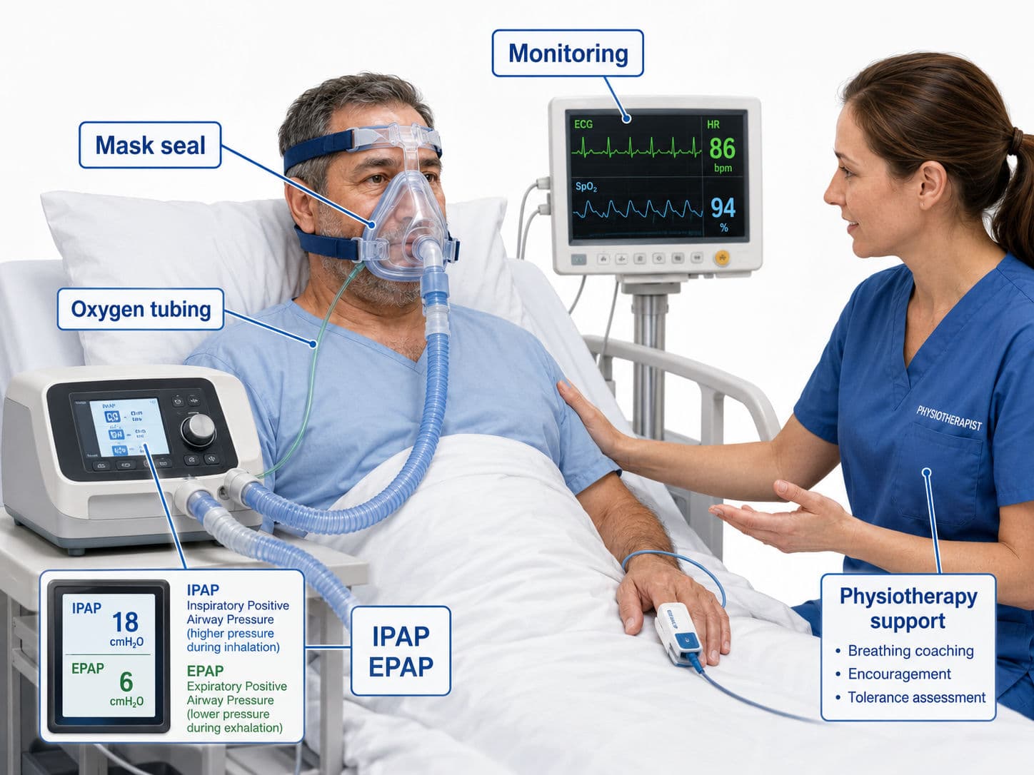
Bilevel positive airway pressure, commonly called BiPAP, provides two pressure levels: a higher inspiratory pressure and a lower expiratory pressure. The higher pressure is called IPAP, and the lower pressure is called EPAP. The difference between IPAP and EPAP is pressure support. This pressure support increases tidal volume, reduces work of breathing and helps remove carbon dioxide. Therefore, BiPAP is especially important in acute hypercapnic respiratory failure, such as acute exacerbation of COPD with respiratory acidosis (Davidson et al., 2016; Rochwerg et al., 2017).

In a nutshell, CPAP mainly recruits alveoli. BiPAP improves ventilation.
Indications for NIV
The most exam-relevant indication for BiPAP is acute exacerbation of COPD with persistent hypercapnic respiratory acidosis. NIV is recommended when the patient remains acidotic despite optimal medical therapy, usually with pH ≤ 7.35 and raised PaCO₂ (Davidson et al., 2016; National Institute for Health and Care Excellence [NICE], 2023; Rochwerg et al., 2017). GOLD also recognises ventilatory support, including NIV, as an important intervention in COPD patients with acute respiratory failure because it improves gas exchange and reduces ventilatory load (Global Initiative for Chronic Obstructive Lung Disease [GOLD], 2026).
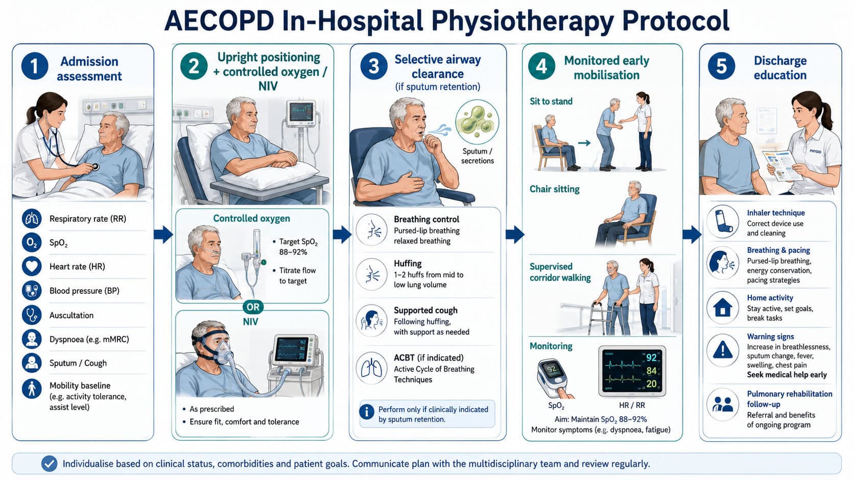
In AECOPD, the patient may present with severe breathlessness, tachypnoea, accessory muscle use, reduced chest expansion, CO₂ retention, fatigue and sometimes drowsiness. For a physiotherapist, the blood gas is important, but it should never be interpreted alone. The whole clinical picture matters: respiratory rate, work of breathing, mental status, cough effectiveness, secretion load, haemodynamic stability and ability to cooperate with the interface.

CPAP is more commonly indicated when the primary problem is oxygenation rather than CO₂ clearance. In cardiogenic pulmonary oedema, positive pressure reduces preload and afterload while improving alveolar recruitment, which can reduce breathlessness and oxygen requirement (Rochwerg et al., 2017). In postoperative patients, CPAP may be used to reduce atelectasis and improve oxygenation when shallow breathing, pain and reduced lung volume limit recovery.
NIV may also be used after extubation in selected high-risk patients. Preventive NIV is more useful in patients at high risk of extubation failure, such as those with COPD, obesity, cardiac disease or prolonged ventilation. However, NIV should not be used to delay reintubation in a patient who is clearly deteriorating after extubation (Rochwerg et al., 2017).
NIV After Extubation
Post-extubation NIV is used mainly as preventive support in patients at high risk of extubation failure, rather than as a late rescue strategy after clear deterioration. High-risk groups include patients with COPD, obesity hypoventilation, cardiac disease, a weak cough, prolonged mechanical ventilation, older age or previous extubation difficulty. In these patients, NIV can reduce respiratory muscle load, support ventilation, improve oxygenation and provide a bridge while the patient transitions from invasive ventilation to spontaneous breathing.
The physiotherapist’s role after extubation is to monitor whether NIV is helping or masking deterioration. Assessment should include respiratory rate, SpO₂, work of breathing, cough strength, secretion load, voice quality, alertness and haemodynamic response. Airway clearance, upright positioning, supported coughing, early sitting and graded mobilisation should be planned around NIV breaks or delivered with NIV support if tolerated. However, if the patient develops worsening acidosis, exhaustion, altered consciousness, severe hypoxaemia or haemodynamic instability, NIV should not delay escalation or reintubation.
Common NIV Settings the Physiotherapist Should Recognise
Physiotherapists may not prescribe NIV settings in many hospitals, but they must understand what the numbers mean. This allows safer treatment planning and better communication with the medical team.

In many acute settings, BiPAP may begin with relatively low pressures, such as IPAP around 10–12 cmH₂O and EPAP around 4–5 cmH₂O, then be titrated according to comfort, respiratory rate, tidal volume, oxygenation and blood gas response (Davidson et al., 2016). If PaCO₂ remains high and the patient is tolerating NIV, IPAP may be increased to improve ventilation. If oxygenation is poor, EPAP or oxygen delivery may be adjusted, but clinicians must remember that increasing EPAP without increasing IPAP reduces pressure support.
In acute hypercapnic respiratory failure, oxygen should be controlled. BTS/ICS guidance recommends a target oxygen saturation of 88–92% in acute hypercapnic respiratory failure treated with NIV (Davidson et al., 2016). This is highly exam-relevant because excessive oxygen in CO₂ retainers can worsen hypercapnia.
Physiotherapy Role During NIV
The physiotherapist’s role starts with assessment. Before treatment, check diagnosis, indication for NIV, recent ABG, respiratory rate, SpO₂, heart rate, blood pressure, mental status, secretion load, cough strength, chest expansion and patient tolerance. NIV should not make physiotherapy passive. Instead, it should create a safer window for breathing control, positioning, secretion clearance and graded movement (Frownfelter et al., 2022; Hillegass, 2022).
Positioning is often the first intervention. High sitting or forward-supported sitting improves diaphragmatic efficiency, reduces work of breathing and improves ventilation distribution. In anxious patients, explanation and coaching are essential. A patient fighting the mask may worsen leaks, dyssynchrony and respiratory distress.
Skin protection is another important responsibility. The nasal bridge, cheeks and forehead are common pressure areas. The mask should be snug enough to prevent excessive leak but not so tight that it causes pressure injury. Regular skin checks, correct mask size, protective dressings and planned breaks help prevent NIV-related skin damage.

Secretion clearance must be individualised. NIV is not a replacement for cough. If sputum is retained, physiotherapy may include humidification review, supported huffing, active cycle of breathing techniques, positioning, PEP or OPEP where appropriate, and suction support if the patient cannot clear secretions independently. In patients who desaturate or fatigue quickly, airway clearance may be performed in short planned NIV breaks or with NIV support, depending on tolerance and local practice (Bott et al., 2009; Frownfelter et al., 2022).
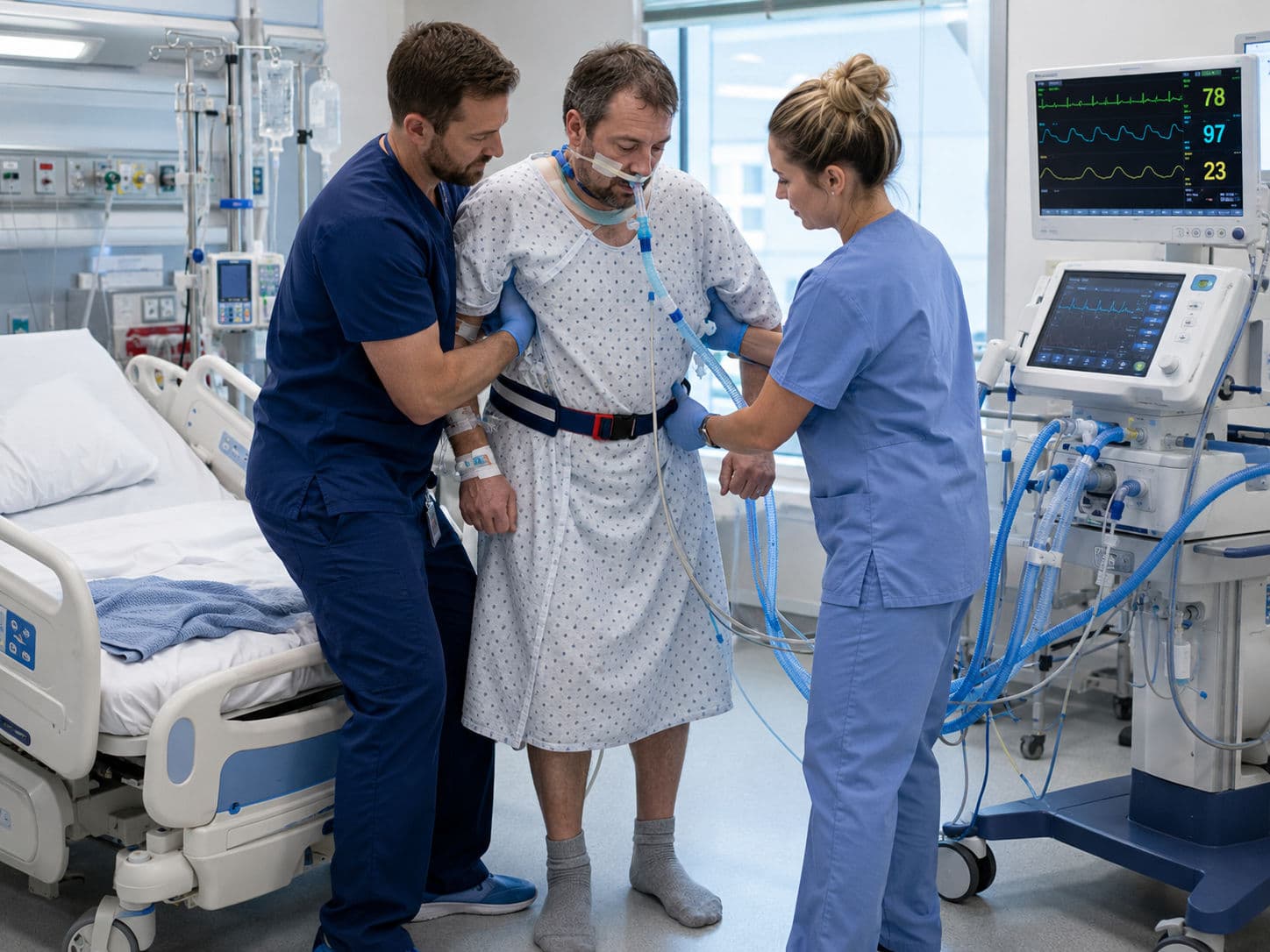
Mobilisation on NIV
A patient being on NIV does not automatically mean bed rest. Mobilisation depends on clinical readiness. The patient should be alert, cooperative, haemodynamically stable, adequately oxygenated, able to tolerate the interface and not showing signs of NIV failure.
Early activity may begin with bed mobility, upright positioning and sitting over the edge of the bed. It can progress to chair sitting, standing, marching on the spot and short walking if the patient remains stable. During mobilisation, the physiotherapist should monitor respiratory rate, SpO₂, heart rate, blood pressure, dyspnoea, fatigue, mask leak and patient comfort.
The safest exam phrasing is 'mobilise during NIV when NIV is stabilising the patient, not when the patient is failing NIV'. If mobilisation causes worsening distress, falling saturation, severe tachycardia, hypotension, reduced consciousness or inability to synchronise with the ventilator, activity should stop and the medical team should be informed.
Weaning from NIV
NIV weaning begins when the underlying reason for NIV is improving. In AECOPD, this usually means improved pH, stable or falling PaCO₂, reduced respiratory rate, reduced accessory muscle activity, improved alertness, manageable secretions and stable oxygen requirement (Davidson et al., 2016).
Weaning can be done in different ways. Some patients first tolerate short NIV-free breaks for meals, communication, airway clearance and mobilisation. Others move from continuous use to daytime breaks and nocturnal NIV. Pressure support may also be reduced gradually if ventilation remains stable. There is no single method that suits every patient, so weaning should be guided by clinical response and ABG trends.
Physiotherapy during weaning should focus on making NIV-free time functional. This includes upright sitting, breathing control, airway clearance, walking practice, ADL training and education. Signs of weaning failure include rising respiratory rate, increasing breathlessness, falling SpO₂, tachycardia, drowsiness, poor cough, exhaustion or worsening acidosis.
Viva-Ready Summary
NIV provides positive pressure support without intubation. CPAP gives one continuous pressure and mainly improves oxygenation by recruiting alveoli. BiPAP gives two pressures: IPAP and EPAP. The pressure support between them improves tidal volume, unloads respiratory muscles and helps clear CO₂. BiPAP is strongly indicated in AECOPD when hypercapnic acidosis persists despite optimal medical treatment. The physiotherapist’s role includes assessment, positioning, breathing control, skin checks, secretion clearance, mobilisation and weaning support.
One-line recall point:
CPAP recruits, BiPAP ventilates, and physiotherapy makes NIV safe, tolerable and functional.
References
Bott, J., Blumenthal, S., Buxton, M., Ellum, S., Falconer, C., Garrod, R., Harvey, A., Hughes, T., Lincoln, M., Mikelsons, C., Potter, C., Pryor, J., Rimington, L., Sinfield, F., Thompson, C., Vaughn, P., & White, J. (2009). Guidelines for the physiotherapy management of the adult medical, spontaneously breathing patient. Thorax, 64(Suppl 1), i1–i51.
Davidson, A. C., Banham, S., Elliott, M., Kennedy, D., Gelder, C., Glossop, A., Church, A. C., Creagh-Brown, B., Dodd, J. W., Felton, T., Foëx, B. A., Mansfield, L., McDonnell, L., Parker, R., Patterson, C. M., Sovani, M., & Thomas, L. (2016). BTS/ICS guideline for the ventilatory management of acute hypercapnic respiratory failure in adults. Thorax, 71(Suppl 2), ii1–ii35.
Frownfelter, D., Dean, E., Stout, M., Kruger, R., & Anthony, J. (2022). Cardiovascular and pulmonary physical therapy: Evidence to practice (6th ed.). Elsevier.
Global Initiative for Chronic Obstructive Lung Disease. (2026). Global strategy for the diagnosis, management, and prevention of chronic obstructive pulmonary disease: 2026 report.
Hillegass, E. A. (2022). Essentials of cardiopulmonary physical therapy (5th ed.). Elsevier.
National Institute for Health and Care Excellence. (2023). Chronic obstructive pulmonary disease in adults: Quality standard QS10, quality statement 7: Non-invasive ventilation.
Rochwerg, B., Brochard, L., Elliott, M. W., Hess, D., Hill, N. S., Nava, S., Navalesi, P., Members of the Steering Committee, Antonelli, M., Brozek, J., Conti, G., Ferrer, M., Guntupalli, K., Jaber, S., Keenan, S., Mancebo, J., Mehta, S., & Raoof, S. (2017). Official ERS/ATS clinical practice guidelines: Noninvasive ventilation for acute respiratory failure. European Respiratory Journal, 50(2), 1602426.