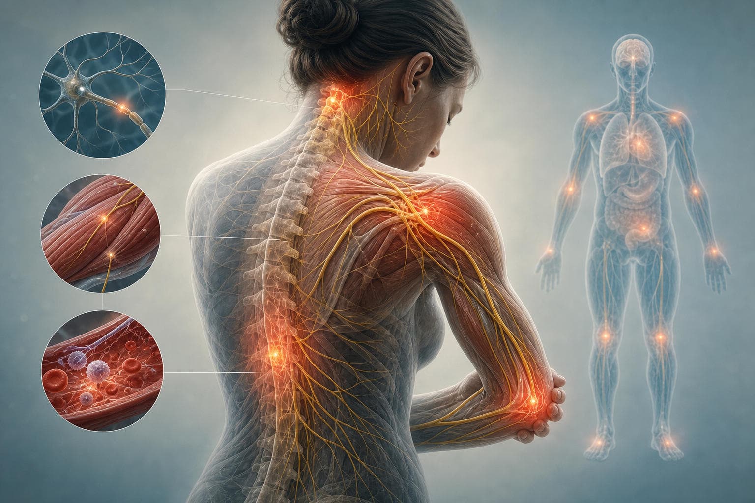
Intercostal Muscles: Anatomy, Function, and Clinical Diagnostic Approaches
Learn the structural anatomy of intercostal muscles, their role in respiration and trunk stability, and a systematic clinical framework for diagnosing rib pain while ruling out life-threatening emergencies.
Every clinical encounter involving chest or thoracic cage discomfort demands rapid, accurate triaging to differentiate benign chest wall conditions from life-threatening cardiopulmonary emergencies. The intercostal muscle group plays a central role in both structural integrity and ventilation, making an injury or dysfunction within this tissue a frequent driver of acute outpatient, urgent care, and emergency department visits. Understanding the structural layers, innervation, and mechanical duties of the intercostal muscle complex enables clinicians to formulate targeted differential diagnoses, order appropriate imaging, and establish clear clinical documentation.
Standardized clinical pathways and EHR documentation templates allow clinical teams to navigate diagnostic ambiguity when patients present with unilateral or bilateral thoracic pain. Implementing structured assessment workflows and precise coding protocols ensures patient safety while optimizing diagnostic precision across care networks.
Intercostal Muscle Anatomy and Layers
The thoracic wall consists of dynamic, layered musculoskeletal units designed to protect underlying vital organs while accommodating continuous volumetric fluctuations during the respiratory cycle. A granular grasp of thoracic wall anatomy is necessary for physical examination localization, procedural safety during thoracic interventions, and structured EHR template design.
The Three Anatomical Layers
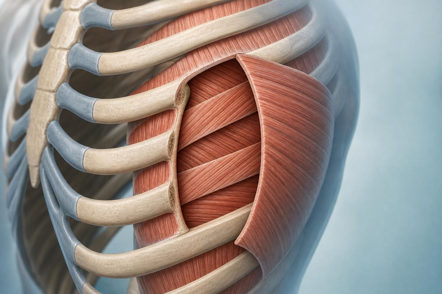
The intercostal space lies between adjacent ribs, spanning eleven distinct intervals on each lateral side of the thoracic cage. Within each space reside three discrete muscular layers, classified by depth and fiber orientation:
- External Intercostal Muscles: The most superficial layer. These eleven pairs of muscles originate from the inferior border of the rib above and insert into the superior border of the rib below. Their fibers run obliquely downward and forward in a "hands-in-pockets" orientation. Anteriorly, at the junction with the costal cartilage, the muscular fibers transition into the external intercostal membrane, which extends medially to the sternum.
- Internal Intercostal Muscles: Positioned directly deep to the external layer. These fibers run obliquely downward and backward, roughly perpendicular to the external intercostals. They originate from the floor of the costal groove of the superior rib and insert into the superior border of the inferior rib. Posteriorly, near the angle of the rib, these muscles are replaced by the internal intercostal membrane, extending to the vertebral column.
- Innermost Intercostal Muscles: The deepest muscular layer. Their fiber orientation mirrors that of the internal intercostal group (downward and backward). This layer is variable in thickness and is separated from the internal intercostal layer by the intercostal neurovascular bundle. Deep to the innermost layer lies the endothoracic fascia, which directly contacts the parietal pleura.
Neurovascular Bundle Organization
A critical clinical landmark within each intercostal space is the subcostal neurovascular bundle. Located in the costal groove along the inferior, internal margin of each rib, the structures are systematically arranged from superior to inferior in the classic "VAN" sequence:
- Intercostal Vein: Occupies the most superior position within the costal groove.
- Intercostal Artery: Positioned intermediate to the vein and nerve, originating posteriorly from the thoracic aorta and anteriorly from the internal thoracic artery.
- Intercostal Nerve: The most inferior structure in the groove, representing the anterior ramus of thoracic spinal nerves (T1 to T11).
Because the intercostal nerve is the most exposed structure in the triad, the inferior rib border is particularly vulnerable to compressive trauma, needle strike during thoracentesis, or traction injury during hyperextension. A collateral neurovascular bundle also runs along the superior border of the inferior rib, though with smaller branch profiles.
Procedures should consistently target the superior border of the lower rib to avoid the main neurovascular tract.
Function: Respiration, Stability, and Movement
The intercostal muscle groups serve two distinct, synchronized functions: modulation of respiratory muscle function and dynamic stabilization of the upper torso during multidirectional kinetic activities.
Respiratory Mechanics: Inspiration vs. Expiration
Ventilation relies on continuous changes in intrathoracic volume, creating pressure gradients that drive air into and out of the lungs. The intercostal muscles act as primary effectors in these volume modifications.
During quiet inspiration, the diaphragm acts as the primary driver of negative intrathoracic pressure. The external intercostal muscles contribute by elevating the ribs. Due to the orientation of the costovertebral and costotransverse joints, rib elevation produces two distinct dimensional changes:
- Bucket-handle movement: Elevation of the middle shafts of the lower ribs increases the lateral (transverse) diameter of the thoracic cavity.
- Pump-handle movement: Elevation of the anterior portions of the upper ribs lifts the sternum forward and upward, expanding the anteroposterior diameter of the chest.
During forced or labored inspiration, such as during strenuous exercise or respiratory distress, external intercostals work synergistically with accessory muscles (sternocleidomastoid, scalenes, and pectoralis minor) to maximize chest volume expansion.
Quiet expiration is primarily a passive process driven by the elastic recoil of lung tissue and the chest wall. However, active or forced expiration (such as coughing, sneezing, or athletic exertion) requires contraction of the internal intercostals and the innermost intercostal group. These muscles depress the ribs, pulling them downward and inward, which actively reduces intrathoracic volume and assists the abdominal wall musculature in forcing air from the lungs.
Torso Stability and Kinetic Chain Integration
Beyond respiration, the intercostal muscles are key components of the core muscular cylinder. They link adjacent ribs together, transforming individual osseous segments into a coherent, semi-rigid cage capable of transferring mechanical loads.
During twisting, lateral flexion, and rotational movements, the intercostal muscles contract isometrically and eccentrically to prevent excessive rib displacement and protect underlying neurovascular structures. In rotational athletics (such as cricket bowling, golf, throwing, and rowing), the intercostals coordinate with the external and internal obliques, serratus anterior, and latissimus dorsi to transmit power from the lower extremities and pelvis through the trunk to the upper extremities. A weakness or mechanical breakdown anywhere along this myofascial chain significantly increases focal stress on the intercostal fibers.
Etiology of Rib Pain: Common Clinical Presentations
Chest wall pain accounts for a substantial proportion of primary care, sports clinic, and emergency visits. Identifying the specific etiology of rib pain causes requires classifying the presentation by onset mechanism, tissue depth, and biomechanical stress patterns.
Acute Mechanical Trauma and Strain
An acute intercostal muscle strain occurs when the muscle fibers stretch beyond their physiological limit or experience a sudden, forceful eccentric load. Common mechanisms include:
- Sudden, extreme torso twisting or lateral bending (e.g., dynamic swings in tennis, batting, or golf).
- Direct blunt-force trauma to the chest wall (e.g., motor vehicle collisions, contact sports, or falls), resulting in direct contusion and fiber disruption.
- Violent or repetitive coughing spasms (e.g., acute bronchitis, pertussis, or exacerbations of chronic obstructive pulmonary disease).
- Heavy lifting with poor biomechanics, particularly overhead pressing or asymmetric carrying tasks.
Strains are clinically graded on a standard 3-tier scale:
- Grade I (Mild): Stretching of a small number of muscle fibers without significant structural disruption; localized tenderness with minimal limitation of respiration or range of motion.
- Grade II (Moderate): Partial tearing of muscle fibers; moderate to severe localized pain, swelling, and palpable tenderness, often causing pain with deep inspiration (splinting).
- Grade III (Severe): Complete rupture of the intercostal muscle fibers, sometimes accompanied by avulsion from the rib margin; marked chest wall instability, severe localized hematoma, and significant respiratory compromise due to splinting.
Chronic Overuse and Repetitive Microtrauma
Chronic or insidious-onset musculoskeletal rib pain typically stems from sustained, submaximal mechanical loading without adequate tissue recovery:
- Sustained postural loading: Prolonged kyphotic or slouched desk posture causes chronic eccentric loading of the posterior intercostals and shortening of anterior thoracic soft tissues.
- Repetitive occupational motions: Jobs requiring continuous reaching, twisting, or overhead loading.
- Asymmetric athletic demands: High-volume rowers, swimmers, and pitchers can develop chronic myofascial trigger points and micro-tears within specific intercostal spaces.
Neuropathic and Radicular Presentations
Thoracic pain may originate from direct nerve irritation rather than primary myofascial tissue damage. An accurate intercostal neuralgia diagnosis is essential when patients present with sharp, burning, lancinating, or dysesthetic pain that travels along a defined dermatomal band corresponding to a specific intercostal nerve. Common triggers include:
- Post-herpetic neuralgia: Latent reactivation of the varicella-zoster virus causing inflammation of the dorsal root ganglion and intercostal nerve trunk.
- Thoracic nerve entrapment: Compression of the intercostal nerve as it pierces the muscular layers or fascia, often near the anterior cutaneous branch termination.
- Thoracic radiculopathy: Compression or irritation of thoracic spinal nerve roots by a herniated disc, osteophyte, or spinal canal stenosis.
Diagnostic Framework for Clinical Practice
Establishing a structured diagnostic framework ensures that clinicians rule out high-acuity systemic conditions first, followed by systematic localization of the chest wall lesion. Implementing structured EHR assessment templates for orthopedic injury streamlines this diagnostic sequence and supports consistent clinical documentation.
Chest Wall / Rib Pain Encounter
- Rule Out Life-Threatening Emergencies (ECG, Vitals, Troponin, D-Dimer if indicated). If unstable: Initiate Acute Resuscitation Pathway.
- Targeted History (Onset, provocative movements, respiratory triggers, trauma).
- Structured Physical Examination
- Inspection (Asymmetry, bruising, rash, respiratory depth)
- Palpation (Focal intercostal space tenderness vs. costochondral junction)
- Provocative Movement (Passive stretch, active contraction, deep inspiration)
- Decision Rules for Diagnostic Imaging (Ottawa Rib Rules / ACR Appropriateness)
- Plain Radiographs (Rule out pneumothorax, displaced rib fractures)
- High-Resolution Ultrasound (Myofascial tear, hematoma, subtle fracture)
- CT / MRI (Complex trauma, diagnostic ambiguity, deep soft-tissue pathology)
- Clinical Management & Coding Classification (ICD-10 M79 / S29 / R07).
Targeted Clinical History
A focused history should isolate the precise onset and characteristics of the pain:
- Onset: Did symptoms begin abruptly following a distinct mechanical event (such as a forceful swing or fall) or develop insidiously over several weeks?
- Pain Quality: Is the pain described as a dull, muscular ache (musculoskeletal), a sharp, pleuritic stitch provoked by inspiration (pleural or intercostal), or a burning, electric sensation traveling around the flank (neuropathic)?
- Modifying Factors: Does pain worsen with specific torso movements, coughing, deep inhalation, or direct pressure on the chest wall? True musculoskeletal rib pain typically worsens with both palpation and torso motion.
- Associated Symptoms: Screen actively for fever, chills, productive cough, hemoptysis, unexplained weight loss, shortness of breath, or cardiac-sounding pressure.
Physical Examination Maneuvers
The physical examination should confirm structural localization while assessing the degree of functional impairment:
- Visual Inspection: Evaluate chest excursion for symmetrical expansion. Look for signs of respiratory splinting, bruising, ecchymosis, soft-tissue swelling, or dermatomal vesicular lesions indicative of herpes zoster.
- Direct Intercostal Palpation: With the patient seated comfortably, palpate systematically along the intercostal spaces from posterior to anterior. Apply firm, direct pressure into the space between the ribs rather than over the osseous rib itself. Focal tenderness localized strictly between ribs strongly points to an intercostal muscle strain.
- Rib Compression / Springing Test: Apply gentle anteroposterior pressure to the sternum while supporting the thoracic spine. Pain provoked along the lateral rib cage suggests an underlying rib fracture rather than an isolated soft-tissue strain.
- Active and Passive Range of Motion:
- Ipsilateral Torso Flexion: Compresses the affected intercostal space, often triggering focal ache or cramping.
- Contralateral Torso Flexion: Stretches the involved intercostal muscle fibers, typically reproducing sharp, localized pain.
- Active Torso Rotation: Evaluates muscle activation under dynamic load.
- Respiratory Provocation: Instruct the patient to take a maximum deep inspiration. Pain localized to the palpated muscle site during full expansion reflects stretch on the external intercostals and chest wall excursion.
Imaging Modalities and Decision Rules
Imaging should not be ordered indiscriminately for straightforward, low-grade chest wall strains. Clinicians should apply clear imaging decision frameworks:
| Imaging Modality | Clinical Indications | Strengths | Diagnostic Limitations |
|---|---|---|---|
| Plain Radiographs (Chest / Rib Series) | High-energy blunt trauma, suspected pneumothorax, suspicion of displaced rib fracture, or ruling out pneumonia. | Fast, widely accessible, low radiation dose; rules out gross osseous and parenchymal emergencies. | Poor sensitivity for non-displaced fractures; cannot visualize soft-tissue intercostal muscle tears. |
| High-Resolution Musculoskeletal Ultrasound | Focal, localized chest wall swelling; suspected intercostal tear or hematoma; identification of non-displaced rib fractures. | Dynamic real-time imaging, zero ionizing radiation, directly visualizes fiber disruption, hematomas, and cortical steps. | Highly operator-dependent; limited penetration in obese patients or beneath the scapula. |
| Computed Tomography (Thoracic CT) | Major polytrauma, suspected occult osseous lesions, non-healing fractures, or evaluating internal visceral involvement. | Comprehensive cross-sectional assessment of osseous, pleural, and mediastinal structures. | Radiation burden; higher cost; generally unnecessary for isolated, uncomplicated soft-tissue strains. |
| Magnetic Resonance Imaging (Thoracic MRI) | Refractory intercostal neuralgia diagnosis, suspected soft-tissue mass, or radiculopathy with spinal cord involvement. | Exceptional soft-tissue contrast; detects bone marrow edema, nerve root impingement, and deep fascial tears. | High cost, longer acquisition time, limited emergency availability. |
Differential Diagnosis and Red Flags
Because thoracic wall pain frequently mimics life-threatening systemic emergencies, clinicians must maintain a structured differential diagnosis for chest and rib pain before confirming a primary musculoskeletal diagnosis.
Life-Threatening Cardiopulmonary Differentials
The following conditions require immediate clinical exclusion prior to managing thoracic pain as a simple intercostal issue:
- Acute Coronary Syndrome (ACS): May present with atypical or pleuritic chest discomfort, particularly in females, elderly individuals, or patients with diabetes. An immediate 12-lead ECG and cardiac biomarker panel are indicated when risk factors are present.
- Pulmonary Embolism (PE): Typically manifests with sudden pleuritic chest pain, tachypnea, tachycardia, and hypoxia. Wells score and D-dimer testing should guide further CT pulmonary angiography.
- Aortic Dissection: Presents as tearing, severe pain radiating to the interscapular region, accompanied by pulse deficits or blood pressure discrepancies between arms.
- Tension or Simple Pneumothorax: Sudden-onset pleuritic pain with dyspnea, decreased breath sounds, and hyperresonance on the ipsilateral side.
Non-Traumatic Musculoskeletal and Systemic Differentials
Once emergencies are ruled out, differentiate intercostal strain from adjacent musculoskeletal and non-musculoskeletal pathologies:
- Costochondritis: Localized inflammation of the costochondral or chondrosternal junctions. Tenderness is elicited directly over the anterior cartilaginous junction rather than within the lateral intercostal spaces.
- Slipping Rib Syndrome: Hypermobility of the false rib cartilages (ribs 8, 9, and 10), causing subluxation and impingement of the adjacent intercostal nerve. Confirmed clinically via the Hooking Maneuver.
- Pleurisy / Pleural Inflammation: Friction between the parietal and visceral pleura produces a sharp, localized pleuritic pain accompanied by a pleural friction rub on auscultation.
- Pre-Eruptive Herpes Zoster: Severe, burning, unilateral dermatomal pain that precedes the classic vesicular skin eruption by 48 to 72 hours.
- Splenic or Hepatic Pathology: Subdiaphragmatic irritation from splenic hematoma/infarction (left) or acute cholecystitis/hepatitis (right) can refer pain to the lower thoracic cage and intercostal spaces.
Red Flag Indicators for Immediate Escalation
The presence of any of the following clinical findings warrants urgent imaging, laboratory evaluation, or specialist referral:
- Vital sign instability: Tachycardia, hypotension, tachypnea, or oxygen saturation below 95% on room air.
- Systemic "B-symptoms": Unexplained fever, night sweats, rigors, or progressive weight loss.
- Hemodynamic or respiratory compromise: Hemoptysis, syncope, severe dyspnea, or stridor.
- Palpable chest wall deformity: Flail segments, crepitus (subcutaneous emphysema), or expanding pulsatile masses.
- Absence of mechanical reproduction: Chest pain that cannot be provoked, reproduced, or altered by palpation, torso motion, or deep inspiration.
- Progression despite rest: Worsening pain at night or complete absence of improvement after two to three weeks of conservative management.
Clinical Management and Rehabilitation
Managing intercostal muscle injuries requires a phased approach that controls acute symptoms, protects healing myofascial fibers, and restores full respiratory excursion and trunk mobility.
Phase 1: Acute Symptom Control and Relative Rest (Days 1 to 7)
The immediate priority is to reduce acute pain, minimize soft-tissue swelling, and prevent shallow breathing complications without fully immobilizing the thorax:
- Relative Rest: Avoid aggravating activities, including heavy lifting, overhead reaching, and forceful trunk rotation. Absolute bed rest should be discouraged.
- Cryotherapy: Apply cold packs wrapped in a thin towel to the affected intercostal space for 15 to 20 minutes every 3 to 4 hours during the first 48 to 72 hours to reduce localized inflammation.
- Pharmacotherapy: Short courses of nonsteroidal anti-inflammatory drugs (NSAIDs) or paracetamol help manage acute inflammation and baseline discomfort. In cases involving severe pain, short-acting muscle relaxants may be considered for nighttime symptom relief.
- Breathing Exercises: Instruct patients to perform gentle, hourly diaphragmatic breathing (taking 5 to 10 slow, deep breaths every waking hour). Patients should be educated not to splint excessively, as prolonged shallow breathing increases the risk of atelectasis and secondary pulmonary infection.
- Avoid Rib Binders: Elastic chest wraps or restrictive rib binders are strictly discouraged; while they temporarily ease movement pain, they restrict vital capacity and significantly increase pneumonia risk.
Phase 2: Subacute Mobility and Myofascial Restoration (Weeks 2 to 4)
As resting pain subsides, therapy shifts toward restoring chest wall compliance, thoracic spine mobility, and core neuromuscular control:
- Thermotherapy: Transition to moist heat applications before stretching or gentle activity to improve tissue extensibility and ease muscular guarding.
- Gentle Thoracic Mobility: Introduce pain-free, active-assisted range-of-motion exercises, including seated cat-cow movements, gentle thoracic extension over a foam roller, and pain-free lateral side bends.
- Soft-Tissue Mobilization: Gentle myofascial release along the surrounding periscapular, latissimus, and pectoral tissues helps resolve secondary compensatory hypertonicity.
- Intercostal Nerve Blocks: For refractory pain or confirmed intercostal neuralgia, a targeted local anesthetic injection (with or without a corticosteroid) along the inferior rib border under ultrasound guidance provides therapeutic relief and facilitates active rehabilitation.
Phase 3: Functional Strengthening and Return to Sport (Weeks 4 to 8+)
The final phase prepares the patient for unrestricted occupational demands or rotational athletic participation:
- Rotational Core Strengthening: Progressive anti-rotation holds (such as Pallof presses), controlled cable chops, and bird-dog exercises to re-establish kinetic chain stability.
- Sport-Specific Load Progression: Progressive re-introduction of swinging, throwing, or heavy lifting drills, starting at 50% velocity and increasing only when completely pain-free across all planes of movement.
- Ergonomic Optimization: Evaluation of workstation setup, postural habits, and equipment fit to eliminate repetitive asymmetric strain on the thoracic wall.
Documentation and Coding Standards
Accurate, standardized medical record entries ensure high-quality patient transitions, support clinical auditing, and prevent reimbursement delays. Health systems should align their EHR clinical forms with standard clinical documentation best practices to capture the necessary specificity.
Essential Documentation Elements
Every clinical note evaluating thoracic wall pain should systematically record:
- Specific anatomical location: Laterality (left, right, bilateral), rib level (e.g., 5th to 6th intercostal space), and line orientation (mid-clavicular, anterior axillary, posterior scapular).
- Mechanism of injury: Detailed narrative describing whether the condition arose from an acute trauma event, dynamic sports exertion, chronic postural strain, or severe coughing fits.
- Objective examination findings: Precise documentation of localized palpation tenderness, provocative movement results (ipsilateral/contralateral side-bending), breath sound auscultation, and presence or absence of chest wall instability or ecchymosis.
- Diagnostic justification: Rationale for obtaining or deferring diagnostic imaging (e.g., meeting criteria under clinical decision rules), and documented exclusion of acute cardiopulmonary red flags.
- Functional impairment: Documented impact on daily activities, work capacity, and spirometry or respiratory depth metrics where applicable.
ICD-10 Coding Matrix
Proper selection of diagnostic codes requires distinguishing between acute traumatic injuries, non-traumatic muscle conditions, and localized pain syndromes. Clinicians and coding teams should consult standard guidance on ICD-10 coding for musculoskeletal conditions to ensure compliance.
| ICD-10 Code | Official Descriptor | Clinical Documentation Requirements |
|---|---|---|
| S29.011A | Strain of muscle and tendon of front wall of thorax, initial encounter | Traumatic acute strain of anterior intercostal/pectoral muscle. Requires specified laterality and encounter type (A = initial, D = subsequent, S = sequela). |
| S29.012A | Strain of muscle and tendon of back wall of thorax, initial encounter | Traumatic strain localized to posterior thoracic wall musculature. Requires initial encounter designation and laterality. |
| M79.1 | Myalgia | Non-traumatic intercostal muscle pain or chronic myofascial pain syndrome without distinct acute trauma history. |
| M79.2 | Neuralgia and neuritis, unspecified | Confirmed intercostal nerve irritation, nerve entrapment, or neuropathic symptoms along the intercostal tract. |
| R07.82 | Intercostal pain | Localized non-cardiac intercostal pain when definitive underlying pathology (muscle vs. nerve) remains undetermined. |
| R07.89 | Other chest pain | Chest wall discomfort, musculoskeletal chest pain, or anterior rib pain not classified under a more specific code. |
| R07.9 | Chest pain, unspecified | Provisional symptom code reserved strictly for triage prior to definitive diagnostic evaluation. |
Frequently Asked Questions
How long does intercostal muscle strain take to heal?
A mild (Grade I) strain typically resolves within 2 to 3 weeks with conservative management, whereas moderate to severe (Grade II or III) tears can require 6 to 8 weeks or longer to regain full functional capacity.
Is imaging necessary for suspected intercostal muscle injury?
Routine imaging is not necessary for mild, non-traumatic presentations; however, plain radiographs or high-resolution ultrasound should be ordered when there is a history of significant blunt trauma, suspected rib fracture, or failure to improve after conservative therapy.
Can intercostal muscle pain affect breathing patterns?
Yes, localized pain during rib cage expansion often causes patients to take shallow, rapid breaths (respiratory splinting), which can lead to alveolar collapse and increase the risk of atelectasis or secondary lung infections.
What exercises are safe during intercostal muscle recovery?
Early in recovery, safe exercises include gentle diaphragmatic breathing, supported thoracic extension, and walking; rotational trunk exercises, heavy overhead lifting, and high-velocity athletic movements should be avoided until basic movements are completely pain-free.
What is the best pain management approach for rib pain?
An effective approach combines short-term oral analgesics or NSAIDs with local cold packs in the acute phase, transitioning to moist heat, gentle myofascial stretching, and, when indicated, targeted intercostal nerve blocks for persistent pain.