Lateral Knee Pain: Assessment & Hip-Stabilization Program
Lateral knee pain, often caused by IT band syndrome and hip weakness, disrupts running, cycling, and daily movement. Learn how to assess your condition and strengthen your hips to resolve outer knee discomfort.

Experiencing pain on outside of knee tissues can disrupt everyday movements like descending stairs, getting out of a chair, or finishing a morning jog. This symptom, clinically known as lateral knee pain, affects runners, cyclists, field athletes, and active individuals alike. The knee sits between the hip and the foot, meaning that pain felt on the outer edge of the joint often stems from mechanical instability higher up the kinetic chain.
Understanding the source of your outer knee discomfort helps you take control of your recovery. While the irritation feels centered at the joint line, the true mechanical driver is frequently weak hip musculature that allows the thigh bone to collapse inward during movement. By evaluating the joint systematically and addressing muscular imbalances with targeted hip-strengthening exercises, you can resolve symptoms and restore healthy joint mechanics.
What Causes Pain on the Outside of Your Knee?
Lateral knee pain can arise from tendons, ligaments, cartilage, or mechanical maltracking. Distinguishing between these structures guides the appropriate rehabilitation plan.
| Potential Cause | Primary Location | Key Distinguishing Feature |
|---|---|---|
| IT Band Syndrome | 1 to 2 cm above lateral joint line | Burning ache, aggravated by downhill walking or running |
| Lateral Meniscus Tear | Outer joint line | Deep catching, clicking, or swelling after twisting |
| LCL Sprain | Lateral collateral ligament path | Tenderness after a direct blow or awkward twist |
| Patellofemoral Pain | Lateral aspect of kneecap | Grinding or aching during deep squats and stair descent |
Iliotibial Band Syndrome
Iliotibial band syndrome (IT band syndrome) is the most frequent cause of outer knee discomfort in active individuals.
The iliotibial band is a thick band of connective tissue running along the outside of the thigh from the pelvis down to the tibia. During repetitive bending and straightening of the knee, the posterior fibers of the band compress the fat pad and tissue overlying the lateral femoral epicondyle, producing sharp or burning iliotibial band pain.
Lateral Meniscal Pathology
The lateral meniscus acts as a fibrocartilage shock absorber on the outer side of the knee joint. A tear can develop gradually from repetitive loading or suddenly from a sharp pivoting motion. Symptoms include joint-line tenderness, delayed swelling, and mechanical symptoms like locking or catching.
Lateral Collateral Ligament Sprains
The lateral collateral ligament (LCL) runs along the outside of the knee, stabilizing it against outward bowing forces (varus stress). LCL sprains usually follow a direct impact to the inside of the knee or a severe twisting motion, resulting in pinpoint tenderness, localized swelling, and joint instability.
Patellofemoral Pain Syndrome
When the patella tracks laterally within the femoral groove, pressure concentrates on the lateral facet of the kneecap.
Tight lateral retinacular tissues can refer pain toward the outer compartment of the knee, mimicking tissue friction around the joint line.
Hip Weakness as a Root Mechanism
Hip weakness is the primary biomechanical driver for persistent pain on outside of knee structures. When the gluteus medius and deep external rotators lack strength, the femur drops inward (adducts) and rotates internally during single-leg weight bearing. This dynamic knee valgus pulls the iliotibial band taut across the lateral femoral condyle, magnifying friction and joint compression.
How to Evaluate Lateral Knee Pain at Home

A structured lateral knee assessment helps you narrow down potential causes before starting an exercise routine.
Work through these four assessment steps carefully on a level surface. For a broader diagnostic perspective across other joint structures, review our comprehensive knee evaluation framework.
Step 1: Local Palpation Test
Sit with your knee bent at 90 degrees. Press your thumb along the lateral joint line (the groove between the thigh and shin bone), then move 1 to 2 cm higher onto the bony bump of the lateral femoral epicondyle. Joint line tenderness suggests meniscal or ligament involvement, whereas tenderness slightly above the joint line on the epicondyle indicates IT band irritation.
Step 2: Modified Ober Test
Lie on your unaffected side with your lower knee bent for stability. Have a partner gently support your top leg, bend the knee to 90 degrees, bring the hip back into slight extension, and slowly lower the thigh toward the bed or floor. If your top thigh remains elevated parallel to the table and cannot drop below horizontal without hip rotation, significant lateral fascial tightness is present.
Step 3: Noble Compression Test
Lie on your back. Bend your painful knee to 90 degrees. Apply firm thumb pressure directly over the lateral femoral epicondyle (the outer bony prominence just above the joint line). Slowly straighten your knee while maintaining this pressure. If you experience a sharp reproduction of your typical pain at roughly 30 degrees of knee flexion, the test is positive for IT band friction.
Step 4: Single-Leg Squat Observation
Stand in front of a mirror on the affected leg with your hands on your hips. Slowly perform a shallow single-leg squat.
Watch the path of your kneecap:
- If your knee caves inward past your big toe, hip abductor weakness and dynamic valgus are present.
- If your opposite hip drops downward, your stance-leg gluteus medius is struggling to maintain pelvic alignment.
- If you can maintain the knee directly over the second toe with a level pelvis, your dynamic control is intact.
If you experience severe swelling, an inability to bear weight, or a locked joint that will not straighten, seek an evaluation from an orthopedic doctor or physical therapist before attempting corrective exercises.
Why Hip Strength and IT Band Mobility Matter
The hip and knee function together as a connected kinetic chain. The iliotibial band is not an active muscle; it is a passive fascial structure tensioned by two main muscles at the pelvis: the tensor fasciae latae (TFL) and the gluteus maximus.
When the primary pelvic stabilizer, the gluteus medius, becomes underactive, the body compensates by overworking the TFL. Because the TFL blends directly into the anterior border of the IT band, chronic TFL overactivity increases resting tension throughout the entire lateral fascia.
Gluteus Medius Weakness leads to Compensatory TFL Overactivity, which leads to Increased IT Band Tension and Inward Knee Collapse (Valgus), which leads to Heightened Friction at Lateral Femoral Epicondyle, which leads to Pain on Outside of Knee.
Strengthening the posterior hip musculature, specifically the gluteus medius and gluteus maximus, offloads the TFL and restores neutral lower-limb alignment during walking, stair navigation, and running.
Essential Hip-Strengthening Exercises for Lateral Knee Pain
To resolve pain on outside of knee structures, perform targeted hip strengthening exercises 3 to 4 days per week.
These movements build active pelvic stability and prevent dynamic knee valgus. For advanced training progressions, explore our full hip strengthening program.
1. Side-Lying Clamshells
Lie on your side with your hips flexed to 45 degrees and knees bent to 90 degrees. Keep your feet pressed together.
Starting Position: Hips at 45 degrees, Knees at 90 degrees, Feet touching
Movement: Rotate top knee upward without rolling pelvis backward
Hold: Pause for 2 seconds at the top
- Target: Gluteus medius and deep external rotators.
- Volume: 3 sets of 15 repetitions per side.
- Form cue: Do not let your pelvis roll backward as you open the knee. Keep your core tight.
2. Side-Lying Hip Abduction
Lie on your unaffected side with your bottom leg bent for support. Keep your top leg completely straight, with the toes pointed slightly downward toward the floor and the heel driving upward.
- Target: Gluteus medius (posterior fibers).
- Volume: 3 sets of 12 to 15 repetitions per side.
- Form cue: Lift your leg slightly behind the midline of your body rather than in front.
3. Glute Bridge with Resistance Band
Place a looped resistance band just above your knees. Lie on your back with your knees bent and feet flat on the floor, hip-width apart. Push outward against the band to keep your knees in line with your shoulders, then drive through your heels to raise your hips.
- Target: Gluteus maximus and hip abductors.
- Volume: 3 sets of 12 repetitions, holding the top position for 3 seconds.
- Form cue: Keep your ribs down and avoid arching your lower back at the top.
4. Single-Leg Stance with Hip Hike (Pelvic Drop)
Stand on a low step on your affected leg, letting your free leg hang off the edge. Keep your stance leg straight without locking the knee. Slowly lower your non-stance hip downward, then contract your stance-side glute to pull the pelvis back up past level.
- Target: Weight-bearing gluteus medius endurance.
- Volume: 3 sets of 10 to 12 repetitions per leg.
- Form cue: Perform the movement slowly with a 2-second lowering phase and a 1-second squeeze at the top.
5. Lateral Band Walks
Place a mini resistance band around your ankles or just above your knees. Assume a quarter-squat position with your feet hip-width apart. Step laterally, maintaining constant tension on the band.
- Target: Dynamic lateral hip stability.
- Volume: 3 sets of 10 to 12 steps in each direction.
- Form cue: Keep your feet pointing straight forward; do not let your feet turn outward as you step.
6. Monster Walks
With the resistance band around your ankles, maintain a quarter-squat position and take diagonal steps forward, stepping wide with each stride. Reverse the movement by stepping backward.
- Target: Combined hip abduction and extension.
- Volume: 3 sets of 10 steps forward and 10 steps backward.
- Form cue: Maintain a wide base throughout the movement so the band never loses tension.
IT Band Mobility and Soft-Tissue Release
Releasing tension in the muscles that anchor into the lateral fascia reduces resting strain across the knee. Avoid aggressive rolling directly over the bony lateral femoral condyle, as this compresses already inflamed tissues. Focus soft-tissue work on the muscular bellies above the joint. For detailed video guides and manual therapy options, view our advanced IT band mobility work.
Foam Rolling the Lateral Hip and Quad
Position a foam roller under your outer hip, targeting the tensor fasciae latae and the lateral aspect of your quadriceps (vastus lateralis). Roll slowly from just below the hip crest down to mid-thigh. Spend 60 to 90 seconds per side, pausing on tender muscular knots for 20 to 30 seconds while taking deep breaths.
Safe Foam Rolling Zones:
- Upper lateral hip (TFL muscle belly)
- Anterolateral thigh (Vastus Lateralis)
- Gluteal complex
Avoid:
- Direct pressure on the lateral knee joint line
- Rolling directly over the lateral femoral epicondyle
Figure-4 Gluteal Stretch
Lie on your back with both knees bent. Cross your affected ankle over the opposite knee. Reach through and grasp behind the thigh of the uncrossed leg, pulling it gently toward your chest until you feel a firm, comfortable stretch in your outer hip and glute. Hold for 30 to 45 seconds, repeating 3 times per side.
Half-Kneeling Hip Flexor and TFL Stretch
Kneel on your affected knee with your opposite foot flat on the floor in front of you. Tuck your pelvis underneath you (posterior pelvic tilt) and gently shift your weight forward until you feel a stretch in the front of your hip. To target the TFL, reach the arm on your kneeling side upward and lean your torso slightly toward the opposite side while rotating your ribcage outward. Hold for 30 seconds; repeat 3 times.
Return-to-Activity Protocol and Prevention
A structured progression allows healing tissues to adapt to mechanical load without triggering recurring pain on outside of knee structures. Progress through each phase only when the prior phase is pain-free. If you are preparing to run again, consult our phased return-to-activity guidelines.
Phase 1: Acute Calming and Mobility (Days 1 to 7)
- Reduce high-impact activities, running, and deep jumping.
- Perform soft-tissue foam rolling to the TFL and quadriceps daily.
- Complete non-weight-bearing hip exercises: side-lying clamshells and side-lying hip abduction (3 sets of 15 repetitions daily).
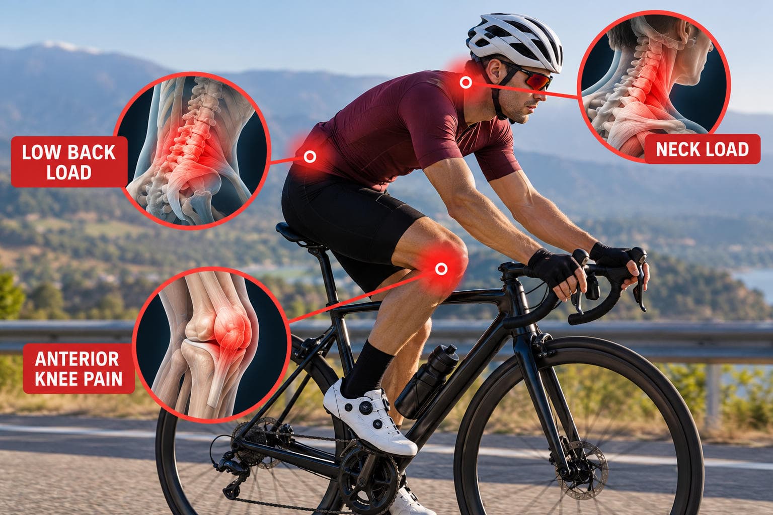
- Maintain cardiovascular fitness with low-resistance stationary cycling (seat raised high to limit knee flexion past 90 degrees) or pool swimming with a gentle flutter kick.
Phase 2: Static and Dynamic Strength (Weeks 2 to 3)
- Introduce loaded standing exercises: glute bridges with resistance bands, pelvic drops, and lateral band walks.
- Begin eccentric quadriceps and hamstring work, such as slow tempo split squats, keeping the knee aligned over the second toe.
- Continue soft-tissue release 3 times per week following exercise sessions.
- Begin flat-surface walking intervals (20 to 30 minutes), monitoring for any delayed ache.
Phase 3: Dynamic Loading and Impact Preparation (Weeks 3 to 4)
- Progress to monster walks, single-leg deadlifts, and step-downs from a low platform.
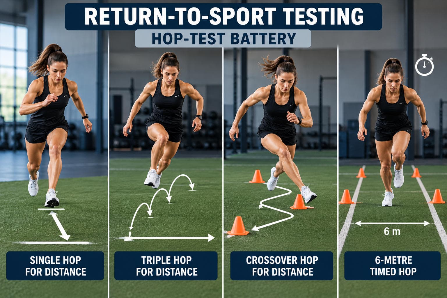
- Add light plyometrics, such as low-amplitude two-foot hops and controlled lateral bounds, emphasizing soft landings without inward knee collapse.
- Perform exercises 3 days per week with progressive resistance bands.
Phase 4: Gradual Sport-Specific Progression (Weeks 4 to 6+)
- Initiate a walk-run interval program on flat, even terrain (for example, 1 minute of jogging alternating with 2 minutes of walking for 20 minutes total).
- Avoid downhill running, cambered road edges, and high-cadence cycling until jogging is entirely symptom-free.
- Maintain a twice-weekly hip-strengthening routine indefinitely to protect against reinjury.
Frequently Asked Questions
Can lateral knee pain go away on its own?
Mild lateral knee pain from minor overuse may settle with rest, but symptoms frequently return once you resume normal activity if underlying hip weakness and movement flaws are not corrected.
Is my condition IT band syndrome or patellofemoral pain?
IT band syndrome typically presents as a localized, sharp or burning pain on the outer bony prominence of the knee, whereas patellofemoral pain is felt as a broader ache around, behind, or under the kneecap during loaded bending.
How often should I do these hip-strengthening exercises?
Perform the core hip-strengthening routine 3 to 4 times per week on non-consecutive days, while soft-tissue foam rolling and mobility stretches can be done daily.
When should I see a doctor for lateral knee pain?
You should see a doctor promptly if you notice rapid joint swelling, cannot bear weight on the leg, experience joint locking or catching, or have pain that persists after 2 weeks of conservative self-care.
Can tight hamstrings cause outside knee pain?
Yes, tightness in the lateral hamstring (biceps femoris) can place tension near the head of the fibula and alter knee mechanics, contributing to pain on the outer side of the joint.