Orbicularis Muscle: Anatomy and Nerve Supply
The orbicularis oculi and orbicularis oris are concentric sphincter muscles essential for eye closure, lacrimal drainage, and oral competence. Understanding their anatomy, fiber organization, and facial nerve innervation is vital for clinicians managing facial nerve pathology and dysfunction.
Overview of the Orbicularis Muscle
The term orbicularis refers to striated muscles arranged in concentric rings or ellipses around the natural apertures of the face. Unlike standard skeletal muscles that span between fixed skeletal landmarks to mobilize synovial joints, the orbicularis muscle groups integrate deeply with adjacent soft tissues, dermis, and neighboring mimetic muscles. They act primarily as biological valves that constrict, compress, and regulate facial orifices.
Within specialized facial muscle anatomy, the two primary orbicularis muscles are:
- Orbicularis oculi: Encircles the orbit, eyelids, and periorbital margins. It mediates voluntary forced closure, gentle reflex blinking, and the active pumping of the lacrimal drainage system.
- Orbicularis oris: Encircles the oral aperture within the upper and lower lips. It coordinates oral seal, lip protrusion, puckering, sucking, and the precise phonation of bilabial consonants (such as /p/, /b/, and /m/).
Both muscles develop embryologically from the second pharyngeal arch during gestational weeks six to eight. Consequently, they share a common motor innervation via the terminal branches of Cranial Nerve VII (the facial nerve). Despite this common origin, the functional subdivisions, architectural stratification, and terminal axonal densities of each muscle differ substantially to accommodate their distinct physiological tasks.
Orbicularis Oculi: Anatomy and Subdivisions
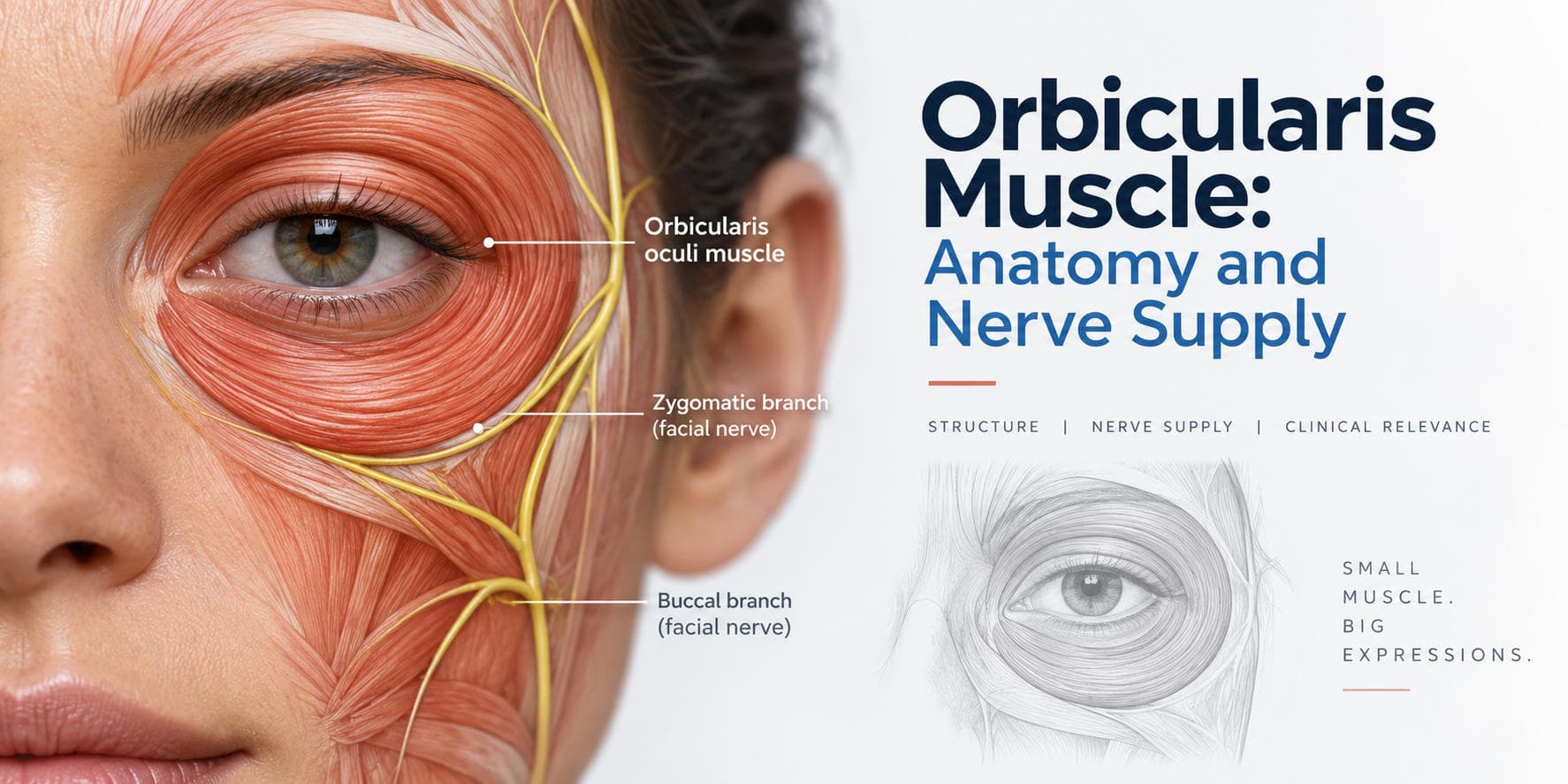
The orbicularis oculi anatomy consists of a broad, thin, elliptical sheet of striated muscle resting immediately deep to the periorbital subcutaneous tissue. Anatomists and surgeons divide it into three structurally distinct parts based on location, fiber size, and physiological role: the orbital, palpebral, and lacrimal portions.
| Sub-portion | Anatomical Origin | Insertion / Termination | Primary Function |
|---|---|---|---|
| Orbital Portion | Nasal part of frontal bone, frontal process of maxilla, medial palpebral ligament | Fibers loop around orbit, interdigitating with frontalis, corrugator, and zygomaticus muscles | Powerful, voluntary forced eyelid closure; protective squinting |
| Palpebral Portion (Preseptal and Pretarsal) | Medial palpebral ligament and posterior lacrimal crest | Lateral palpebral raphe and lateral canthal tendon | Involuntary reflex blinking, gentle voluntary closure, tear film distribution |
| Lacrimal Portion (Horner's Muscle) | Posterior lacrimal crest and lacrimal fascia | Tarsal plates and lateral palpebral raphe | Compresses lacrimal sac; facilitates active tear drainage |
Orbital Portion
The orbital portion forms the outer peripheral zone of the muscle. It arises from the nasal portion of the frontal bone, the frontal process of the maxilla, and the central band of the medial palpebral ligament. Its broad circular fibers sweep outward over the orbital margin, extending superiorly into the forehead, laterally over the temple, and inferiorly onto the cheek. These fibers have no true bony insertion laterally. Instead, they interlace with adjacent mimetic structures, including the frontalis, corrugator supercilii, and procerus muscles superiorly, and the zygomaticus major and levator labii superioris inferiorly. The orbital segment contains a higher ratio of fast-twitch (Type II) muscle fibers, enabling voluntary closure of the eyes during protective grimacing or exposure to bright light and foreign objects.
Palpebral Portion
The palpebral portion is thin, pale, and situated directly within the eyelids. It is divided anatomically into two zones:
- Preseptal segment: Lies superficial to the orbital septum in both upper and lower lids. Fibers arise from the superficial and deep slips of the medial palpebral ligament and sweep laterally to join at the lateral palpebral raphe.
- Pretarsal segment: Rests directly on the anterior surface of the superior and inferior tarsal plates. Its fibers are firmly anchored medially to the posterior lacrimal crest and lateral palpebral raphe via a common lateral canthal tendon. The ciliary bundle (muscle of Riolan) sits at the eyelid margin, maintaining apposition of the lid against the globe.
The palpebral fibers consist predominantly of fatigue-resistant, slow-twitch (Type I) muscle fibers. This portion controls involuntary blinking reflexes, spontaneous micro-blinks that distribute the precorneal tear film, and gentle voluntary eyelid closure during sleep.
Lacrimal Portion (Horner Muscle)
The lacrimal portion, historically designated as Horner's muscle or the tensor tarsi, represents the deep, posterior component of the pretarsal orbicularis. It originates from the upper posterior lacrimal crest of the lacrimal bone and passes behind the lacrimal sac. As its fibers course laterally, they divide into two slips that surround the superior and inferior canaliculi before blending with the pretarsal fibers of each eyelid. Contraction of the lacrimal portion pulls the eyelid margins medially and exerts traction on the lacrimal diaphragm, generating negative hydrostatic pressure that draws tears from the conjunctival sac through the puncta and into the lacrimal drainage system.
- Orbital (Forced Close)
- Palpebral (Blinking)
- Lacrimal (Tear Pump)
Orbicularis Oris: Anatomy and Structure

The orbicularis oris is not a simple, single sphincter muscle. It is a complex architectural meshwork of intrinsic and extrinsic muscle fibers that encircle the mouth, creating a versatile muscular ring. It occupies the full thickness of the lips, situated between the labial skin externally and the oral mucous membrane internally.
Intrinsic and Extrinsic Fiber Architecture
The architecture of the orbicularis oris comprises two primary layers:
- Deep (Intrinsic) Layer: Consists of circular, intrinsic fibers intrinsic to the lips (pars peripheralis and pars marginalis). The marginal part runs close to the vermilion border and is responsible for inverting the lips and modulating fine movements during speech and whistling. The peripheral part forms the bulk of the lip tissue, drawing the lips against the dental arches.
- Superficial (Extrinsic) Layer: Formed by the convergence and decussation of numerous radiating facial expression muscles at a dense fibrous condensation located just lateral to each oral commissure, termed the modiolus.
Modiolus and Muscular Attachments
The modiolus serves as the central dynamic anchor for the midface and lower face. Multiple muscles insert into and interchange fibers with the orbicularis oris:
- Superiorly: Levator labii superioris, levator labii superioris alaeque nasi, zygomaticus major, zygomaticus minor, and levator anguli oris.
- Laterally: Buccinator, risorius, and platysma fibers.
- Inferiorly: Depressor anguli oris, depressor labii inferioris, and mentalis.
Through this interconnected network, the orbicularis oris provides a diverse range of motor outputs. It can compress the lips against the teeth, protrude the lips forward (puckering), narrow the mouth opening, elevate or depress the labial margins, and maintain an airtight seal during the oral preparatory and oral transport phases of deglutition.
Nerve Supply: Facial Nerve Branching Pattern
Motor control over both orbicularis muscle complexes is provided entirely by Cranial Nerve VII (the facial nerve). The facial motor nucleus is situated in the ventrolateral pontine tegmentum of the brainstem. Its axons course dorsally, loop around the abducens (CN VI) nucleus to form the internal genu, and emerge from the brainstem at the cerebellopontine angle.
- Facial Motor Nucleus (Pons)
- Cerebellopontine Angle to Internal Acoustic Meatus
- Facial Canal (Labyrinthine to Tympanic to Mastoid)
- Stylomastoid Foramen
- Parotid Gland Main Trunk Bifurcation (Pes Anserinus):
- Temporofacial
- Cervicofacial
- Terminal branches::
- Temporal
- Zygomatic
- Buccal
- Marginal Mandibular
- Cervical
After traversing the internal acoustic meatus, the facial canal within the temporal bone, and exiting through the stylomastoid foramen, the main trunk enters the posteromedial surface of the parotid gland. Within the parotid parenchyma, the nerve divides at the pes anserinus into two primary divisions: the superior temporofacial and inferior cervicofacial trunks.
These trunks further divide into five classic terminal branching groups: temporal (frontal) branches, zygomatic branches, buccal branches, marginal mandibular branches, and cervical branches. These branches form extensive terminal anastomotic arcades across the midface, known as the parotid plexus. This network provides a degree of redundancy for certain midfacial mimetic muscles, though terminal branches to peripheral sphincters often retain specialized innervation patterns.
Innervation of Orbicularis Oculi
The orbicularis oculi receives motor input through the superior branches of the facial nerve, primarily the temporal and zygomatic branches, with accessory input to inferior fibers from buccal branches.
| Branch | Target |
|---|---|
| Temporal Branch | Orbital Portion (Superior) and Upper Preseptal |
| Zygomatic Branch | Lower Palpebral (Pretarsal/Preseptal) and Lateral Canthus |
| Buccal Branch | Inferior Lacrimal / Deep Medial Striations |
Temporal Branch Pathway
The temporal (or frontal) branch of the facial nerve exits the superior border of the parotid gland and crosses the superficial surface of the zygomatic arch. It travels within the innominate (subgaleal) fascia, deep to the temporoparietal fascia (superficial temporal fascia). It supplies:
- The superior half of the orbital portion of the orbicularis oculi.
- The upper preseptal palpebral fibers.
- The adjacent frontalis and corrugator supercilii muscles.
Because these motor axons enter the deep muscular surface along its superolateral border, surgical dissection along the temporal region must follow strict anatomical planes to avoid transecting these branches, which leads to brow ptosis and incomplete upper lid excursion.
Zygomatic Branch Pathway
The zygomatic branches traverse horizontally across the zygomatic arch and cheek, passing deep to the zygomaticus major muscle. These branches supply:
- The lateral and inferior segments of the orbital portion.
- The lower eyelid preseptal and pretarsal segments.
- The critical motor units mediating involuntary blinking reflexes.
The rich anastomotic network between the temporal and zygomatic branches in the periorbital zone helps preserve partial blinking even after isolated nerve trauma, though targeted terminal branches to the pretarsal fibers remain essential for functional lid closure.
Lacrimal Innervation Details
Fibers running to the lacrimal portion (Horner's muscle) are innervated by deep terminal twigs from the zygomatic and upper buccal branches that penetrate the medial sub-orbicularis plane. This configuration maintains the lacrimal pump mechanism alongside the rhythmic contraction of the preseptal palpebral fibers.
Innervation of Orbicularis Oris
The orbicularis oris innervation is supplied by the lower branches of the facial nerve: the buccal branches and the marginal mandibular branch.
Buccal Branch Contribution
The buccal branches of CN VII travel anteriorly across the masseter muscle, passing inferior to the parotid duct or closely paralleling its course. They supply:
- The superior half of the orbicularis oris (upper lip fibers).
- The modiolus and midfacial elevators (levator labii superioris, zygomaticus major/minor).
- The buccinator muscle, which directly integrates into the lateral lip margins.
Terminal buccal twigs enter the deep surface of the orbicularis oris muscle fibers near the lateral third of the upper lip, directing the puckering, closure, and elevation components of oral movements.
Marginal Mandibular Branch Contribution
The marginal mandibular branch emerges from the inferior pole of the parotid gland and courses across or slightly inferior to the lower border of the mandible, deep to the platysma and superficial to the facial artery and vein. It then loops upward across the mandibular border to enter the lower lip musculature. It supplies:
- The inferior half of the orbicularis oris (lower lip fibers).
- The depressor labii inferioris, depressor anguli oris, and mentalis muscles.
| Structure | Innervation |
|---|---|
| Upper Lip (Orbicularis Oris Superior) | Buccal Branches (CN VII) |
| Lower Lip (Orbicularis Oris Inferior) | Marginal Mandibular Branch (CN VII) |
| Commissure / Modiolus | Dual Buccal / Mandibular Anastomoses |
Bilateral Innervation and Motor Units
A unique feature of the perioral musculature is the presence of midline fiber decussation and overlapping bilateral innervation. Terminal axonal arborizations from the left and right facial nerves cross the midline slightly within the philtrum and lower labial central zone. This overlapping network ensures that unilateral peripheral lesions cause asymmetrical distortion and commissure drooping rather than complete paralysis of the entire central lip sphincter. The fine motor unit architecture of the perioral muscles resembles that of the extraocular muscles, allowing for complex vowel shaping, consonantal stops, and precise dynamic embouchure control.
Clinical Applications in Facial Nerve Procedures
Given the central functional and aesthetic roles of the orbicularis oculi and orbicularis oris, both sphincters serve as key clinical indicators in neurological, reconstructive, and cosmetic medicine.
| Clinical Presentation | Anatomical Mechanism | Primary Intervention Target |
|---|---|---|
| Bell's Palsy Lagophthalmos | Denervation of temporal and zygomatic CN VII branches | Lubrication, moisture chamber, upper eyelid weight insertion |
| Perioral Asymmetry | Marginal mandibular / buccal branch denervation | Reconstructive reanimation, modiolus re-anchoring |
| Lateral Canthal Rhytids (Crow's Feet) | Hyperdynamic contraction of orbital orbicularis oculi | Botulinum toxin injection into lateral orbital fibers |
| Hyperdynamic Lip Lines (Smoker's Lines) | Repetitive contraction of pars marginalis fibers | Low-dose superficial toxin to perioral vermilion border |
Bell's Palsy and Peripheral Lesions
In acute peripheral facial nerve palsy, such as Bell's palsy assessment protocols identify, paresis of the orbicularis oculi leads to lagophthalmos: an inability to close the eyelid fully. As the patient attempts eye closure, the eyeball rolls upward and slightly outward, exposing the inferior sclera (Bell's phenomenon). This physiological reflex unmasks the underlying orbicularis muscle weakness.
Without appropriate intervention, including artificial lubrication, nighttime taping, or temporary external lid weights, the exposed cornea suffers from punctate epithelial erosions, microbial ulceration, and potential visual loss. Perioral denervation manifests as ipsilateral labial drooping, effacement of the nasolabial fold, difficulty with speech articulation, and loss of oral continence resulting in drooling.
Neurological Localization: UMN vs. LMN
Evaluating orbicularis muscle function assists clinicians in distinguishing upper motor neuron (UMN) from lower motor neuron (LMN) lesions:
- Upper Motor Neuron Lesion (e.g., Cortical Stroke): The portion of the facial motor nucleus that supplies the upper face (forehead and upper orbicularis oculi) receives bilateral corticobulbar input from both cerebral hemispheres. Consequently, a unilateral UMN lesion preserves voluntary and reflex blinking in the upper orbicularis oculi while causing dense contralateral paralysis of the lower face and orbicularis oris.
- Lower Motor Neuron Lesion (e.g., Bell's Palsy, Temporal Bone Fracture): Destroys the common final pathway, resulting in complete ipsilateral paralysis of both the orbicularis oculi (upper face) and the orbicularis oris (lower face).
Neurotoxin Targeting in Aesthetic Medicine
In aesthetic clinical practice, precise neurotoxin targeting uses botulinum toxin type A to modulate hyperdynamic contractions of the orbicularis muscles:
- Lateral Canthal Rhytids ("Crow's Feet"): Produced by chronic, hyperactive contraction of the lateral orbital portion of the orbicularis oculi. Injections are placed into the superficial subcutaneous plane lateral to the orbital rim, avoiding deeper planes to prevent ptosis from toxin diffusion into the levator palpebrae superioris.
- Perioral Rhytids ("Smoker's Lines"): Caused by repetitive contraction of the pars marginalis and pars peripheralis of the orbicularis oris. Micro-aliquots of toxin injected along the vermilion border soften vertical dynamic lines while preserving sufficient tone for oral competence and bilabial speech.
- Blepharospasm and Hemifacial Spasm: Involuntary hyperkinetic spasms of the orbicularis oculi are managed by serial intramuscular neurotoxin injections into the preseptal and orbital fibers, relieving chronic muscle hypertonicity and restoring functional visual fields.
Facial Reanimation and Reconstructive Strategies
When facial nerve continuity is permanently lost following acoustic neuroma resection, trauma, or radical parotidectomy, surgical intervention restores sphincteric competence using specialized facial reanimation techniques:
- Dynamic Corneal Protection: Platinum or gold weight implantation into the upper eyelid supplements gravity-assisted closure, counteracting orbicularis oculi flaccidity. In cases requiring active motor neurotization, clinicians may perform direct nerve transfers, such as masseteric-to-facial or hypoglossal-to-facial nerve anastomoses.
- Perioral Reconstruction: Restoring orbicularis oris function and oral competence involves dynamic free gracilis muscle transfer or local regional muscle transfers (temporalis muscle transfer) anchored directly to the modiolus, restoring symmetrical oral competence and spontaneous smiling dynamics.
Frequently Asked Questions
What is the function of the orbicularis muscle?
The orbicularis oculi closes the eyelids, controls involuntary blinking, and drives lacrimal fluid drainage. The orbicularis oris coordinates the oral seal, lip puckering, speech articulation, and mastication.
Can the orbicularis muscle be paralyzed on one side?
Yes, unilateral facial nerve injury, stroke, or Bell's palsy can paralyze the ipsilateral orbicularis muscle, leading to incomplete eye closure (lagophthalmos) and perioral drooping.
How do clinicians test orbicularis muscle strength?
Clinicians evaluate orbicularis oculi strength by asking the patient to tightly squeeze their eyes shut against manual resistance, and they assess orbicularis oris strength by asking the patient to puff out their cheeks, whistle, or resist lip opening during standard facial nerve testing.
What happens to the orbicularis muscle with facial nerve injury?
Facial nerve injury causes denervation flaccidity, loss of sphincteric tone, lagophthalmos with corneal exposure risk in the eye, and labial drooling with articulation deficits around the mouth.
Why is the orbicularis muscle targeted in aesthetic procedures?
Superficial injections of botulinum toxin into hyperactive orbicularis muscle fibers soften dynamic wrinkles, such as lateral canthal lines (crow's feet) around the eyes and vertical lines around the lips.