Plantar Fasciopathy: Evidence-Based Management
Understand the clinical features that establish the diagnosis, the examination that distinguishes plantar fasciopathy from its mimics, and the management supported by current guidelines.
Plantar heel pain due to plantar fasciopathy is one of the most common causes of foot pain presenting to clinical practice. The condition is often self-limiting over time, but a substantial proportion of patients still have symptoms two years after onset. That persistence underlines the value of effective, evidence-based management rather than reassurance alone. This article describes the clinical features that establish the diagnosis, the examination that distinguishes plantar fasciopathy from its mimics, and the management supported by current guidelines, including the role of high-load strength training.
What Is Plantar Fasciopathy?
Plantar fasciopathy is a load-related disorder of the plantar fascia at or near its origin on the medial calcaneal tubercle. The term fasciopathy is preferred to fasciitis because the underlying tissue change is degenerative rather than primarily inflammatory, in keeping with the understanding of other load-related soft-tissue conditions.
The plantar fascia is a tensioned structure. It supports the medial longitudinal arch and, through the windlass mechanism, is loaded as the toes extend during the propulsive phase of gait. The condition develops when the load exceeds the tissue's capacity, often due to changes in activity, footwear, or weight-bearing. Several factors raise the cumulative load the fascia must tolerate and therefore raise risk:
- Reduced ankle dorsiflexion
- Prolonged standing or walking at work
- Greater body mass
How Does It Present, and How Is It Diagnosed?
The diagnosis is clinical and rests on a characteristic pattern. Patients describe medial plantar heel pain that is typically worst with the first steps after a period of inactivity, such as on rising in the morning or after sitting. It may ease with initial movement, then worsen again with prolonged weight-bearing. Onset frequently follows an increase in weight-bearing activity or a change of footwear.
Examination brings together several findings:
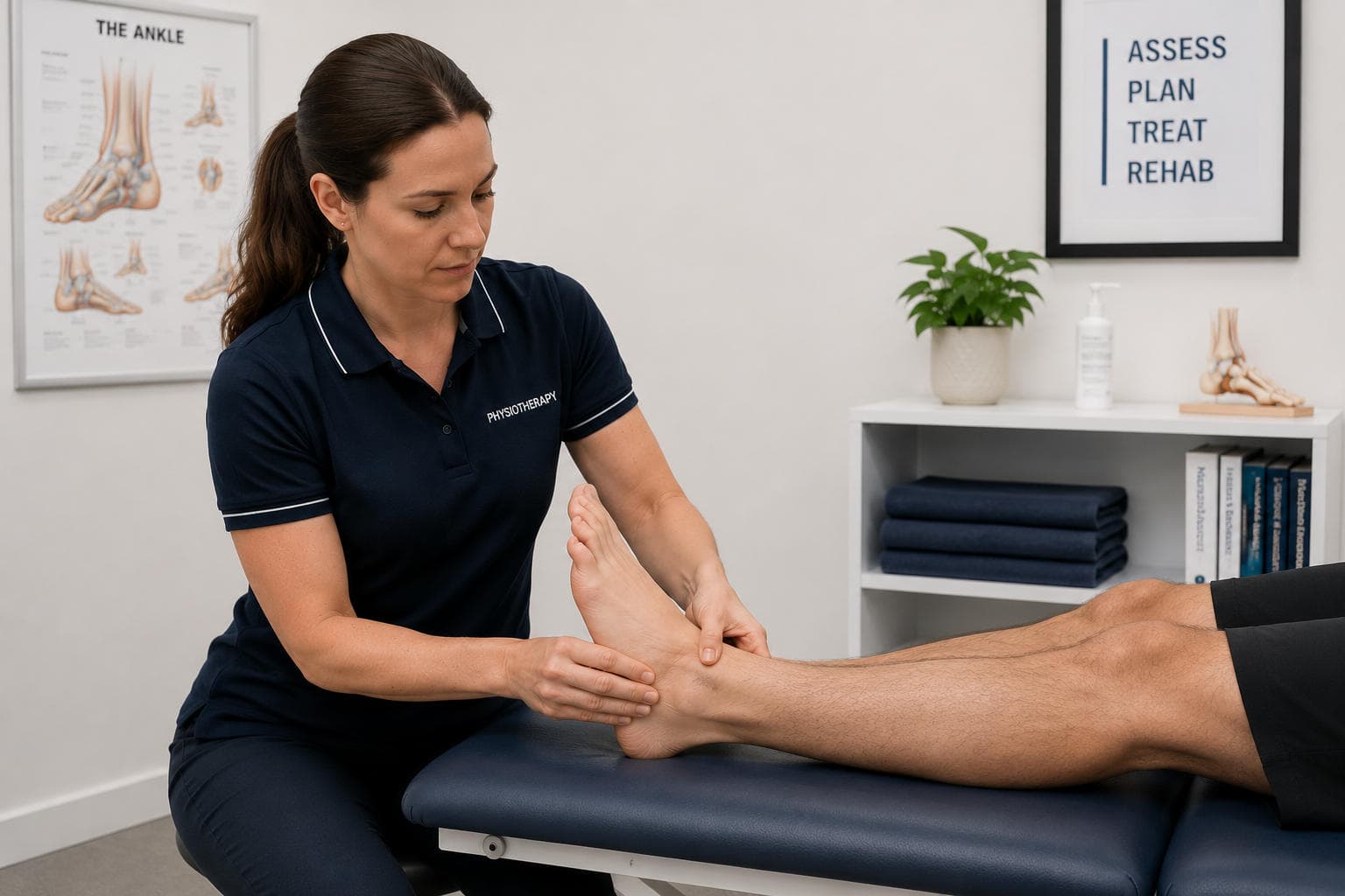
- Tenderness at the proximal plantar fascia insertion on the medial calcaneus
- Ankle dorsiflexion range that may be limited, which increases strain on the fascia
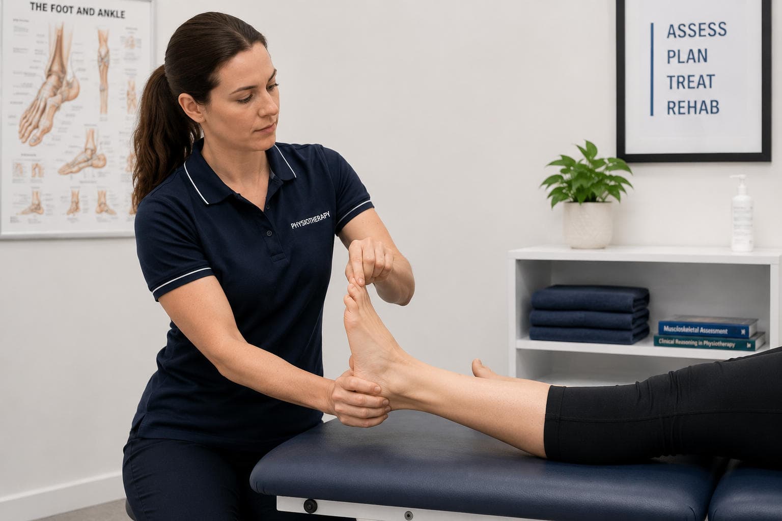
- Positive windlass test, in which extending the toes to tension the fascia reproduces the pain
- Negative tarsal tunnel examination, which helps exclude a neural source.
Together, these establish a working diagnosis without the need for imaging in most cases.
How Is It Distinguished From Other Causes of Heel Pain?
Heel pain has several causes that must be considered before settling on plantar fasciopathy:
- Neural sources, including tarsal tunnel syndrome and entrapment of the nerve to the abductor digiti minimi, may produce burning or radiating symptoms and warrant a neural examination.
- A calcaneal stress fracture should be considered where there is a history of a marked increase in loading and where the calcaneal squeeze reproduces pain.
- A fat pad disorder produces pain more centrally under the heel, rather than at the medial fascial insertion.
- Systemic and inflammatory causes, such as a seronegative spondyloarthropathy, should be considered where the presentation is bilateral, accompanied by other joint symptoms, or otherwise atypical.
The clinical pattern of first-step pain, with localised medial insertional tenderness and a positive windlass test, distinguishes plantar fasciopathy from most of these.
What Does the Evidence Recommend for Management?
Current guidance supports a combination of measures rather than any single treatment:
- Education and load management are foundational: the patient is helped to understand the condition and to modify the loads that provoke it without becoming inactive.
- Stretching of the plantar fascia and of the gastrocnemius-soleus complex is supported.
- Foot orthoses, which may be prefabricated, reduce strain on the fascia.
- Manual therapy to address restricted ankle and foot mobility can be a useful adjunct.
- Taping can provide short-term symptom relief.
Where symptoms are recalcitrant, additional measures fall outside first-line conservative care. The thrust of management is to reduce the provocative load, restore tissue tolerance, and support the patient through a condition whose natural course can be prolonged.
What Is the Role of High-Load Strength Training?
High-load strength training has emerged as a useful loading strategy for plantar fasciopathy. It loads the fascia through the windlass mechanism: the patient performs slow, heavy resistance calf raises with the toes extended over a rolled towel, so dorsiflexion of the toes tensions the fascia as the calf is loaded.
In a randomised trial, a programme of high-load strength training combined with a gel heel insert produced a greater improvement in foot function at three months than plantar-specific stretching with the same insert. The mechanism is consistent with that used for other load-related tendon conditions: progressive, high tensile loading to stimulate adaptation in the tissue.
High-load strength training can be used alongside the other supported measures, and it offers patients a structured, progressive component to their programme. Two caveats keep expectations honest. As with all loading approaches, the benefit accrues over months and depends on consistent performance. By twelve months, there was no significant difference between the strength and stretching groups; therefore, its main value may be a faster initial improvement rather than a better long-term result.
How Does the Windlass Mechanism Inform Treatment?
Understanding the windlass mechanism helps both to explain the condition to the patient and to structure the loading programme. The plantar fascia runs from the calcaneus to the toes. As the toes extend during the push-off phase of gait, the fascia is wound around the metatarsal heads and tensioned, raising the arch and stiffening the foot for propulsion.
This same mechanism is what the windlass test reproduces in the clinic, and it is what high-load strength training deliberately exploits. By performing slow, heavy calf raises with the toes held in extension over a rolled towel, the clinician loads the fascia through windlass-generated tension while simultaneously loading the calf. Framing the fascia as a load-bearing structure that responds to progressive loading — rather than as an inflamed tissue requiring rest — sets the patient's expectations appropriately.
The supported measures work together rather than in isolation:
- Education and load management reduce the provocative loads — often a recent increase in running or standing, or a change of footwear — while preserving activity within tolerance.
- Stretching of the plantar fascia (by extending the toes to recreate the windlass) and of the gastrocnemius-soleus complex addresses the limited ankle dorsiflexion that increases fascial strain.
- Foot orthoses, including prefabricated devices, reduce strain on the fascia and can help particularly in the earlier stages, while taping offers a short-term option.
- Manual therapy directed at restricted ankle and foot mobility can help where stiffness is contributing.
The high-load strength-training protocol itself is straightforward to implement. The patient performs a single-leg heel raise on a step, with a rolled towel under the toes to maintain toe extension, raising slowly over about 3 seconds and lowering over about 3 seconds. Load and repetitions are progressed over time, and the exercise is performed approximately every second day. Because it can be done at home with minimal equipment, it provides the patient with an active, progressive component of their programme. Where symptoms remain recalcitrant despite a well-delivered course of conservative care, additional measures fall outside first-line management and warrant a considered discussion of their evidence and trade-offs rather than early adoption.
When Should Heel Pain Prompt Further Assessment?
Certain features should prompt further assessment rather than continued routine treatment:
- Pain following significant trauma or a sharp increase in loading, suggestive of a stress fracture
- Neurological symptoms suggesting nerve entrapment
- Bilateral heel pain with systemic or inflammatory features
- Failure to respond to a well-delivered course of conservative management
In these situations, imaging, blood investigation, or onward referral may be appropriate to confirm or exclude an alternative diagnosis.
What Is the Prognosis, and What Supports Recovery?
The prognosis is generally favourable, with most patients improving over time, although recovery can be slow and a minority have persistent symptoms. The factors that most support recovery are the patient's understanding of the load-related nature of the problem, their adherence to a progressive loading and stretching programme, and sensible management of footwear and activity.
Setting a realistic timeframe at the outset supports adherence, since a patient who expects rapid resolution may abandon an effective programme prematurely. Equipping the patient to manage load and to continue their exercises independently reduces the likelihood of recurrence. Footwear that supports the foot without being excessively rigid, sensible management of training loads, and maintained calf and foot strength all help to keep symptoms from returning once they have settled, and the patient who understands the load-related nature of the problem is well placed to make these adjustments themselves.
KineticFlow for Plantar Heel Pain
KineticFlow helps you:
- Record the diagnostic pattern: First-step pain, the site of insertional tenderness, dorsiflexion range, and the windlass and tarsal tunnel findings are documented together, so the basis for the diagnosis is explicit.
- Structure the loading programme: Track the progression of high-load strength training and stretching so the current stage and the criteria for advancement are clear.
- Track outcomes against baseline: A foot-specific function score and a pain score are stored at each visit, demonstrating whether the chosen approach is producing meaningful change over time.
- Flag atypical features: Documented screening prompts ensure that features suggestive of a stress fracture, neural entrapment, or an inflammatory cause are not overlooked.
Try KineticFlow for your next patient assessment!
References
https://www.jospt.org/doi/10.2519/jospt.2023.0303
https://doi.org/10.1111/sms.12313